| | | صور طبية هيستولوجى - Histology |  |
| | | كاتب الموضوع | رسالة |
|---|
ابراهيم الشنطي
Admin

عدد المساهمات : 70301
تاريخ التسجيل : 28/01/2013
العمر : 77
الموقع : الاردن
 | | موضوع: صور طبية هيستولوجى - Histology الأحد 18 نوفمبر 2018, 11:56 pm | |
| أولا هيستولوجى - Histology [*] [*] [*] [*] [*] [*] [*] [*] [*] [*] [*] [*] [*] [*] [*] [*] [*] |
| | | | ابراهيم الشنطي
Admin
عدد المساهمات : 70301
تاريخ التسجيل : 28/01/2013
العمر : 77
الموقع : الاردن
| | موضوع: رد: صور طبية هيستولوجى - Histology الإثنين 19 نوفمبر 2018, 12:14 am | |
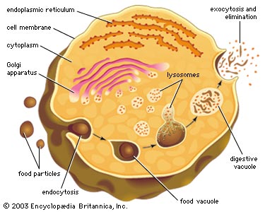
| human cell struture Structure of a typical eukaryotic cell The interior of the cell is divided into the nucleus and the cytoplasm. The nucleus is a spherical or oval shaped structure at the center of the cell. The cytoplasm is the region outside the nucleus that contains cell organelles and cytosol, or cytoplasmic solution. Intracellular fluid is collectively the cytosol and the fluid inside the organelles and nucleus. ــــــــــــــــــــــــــــــــــــــــــــــــــــــــــــــــــــــــــــــــــــــــــــــــــــــــــــــــــــــــــــــــــــــــــــــــــ Membranes are the gateways to the cell. The plasma membrane, is the selective barrier surrounding the cell. It provides a barrier to the movement of molecules between the intra and extracellular fluids. Recall that extracellular means outside the cell. The plasma membrane also serves to anchor adjacent cells together and to the extracellular matrix. Various signals and inputs can alter the sensitivity and permeability of membranes. Cell, or Plasma, membrane - encloses every human cell Structure - 2 primary building blocks include protein (about 60% of the membrane) and lipid, or fat (about 40% of the membrane). The primary lipid is called phospholipid, and molecules of phospholipid form a 'phospholipid bilayer' (two layers of phospholipid molecules). This bilayer forms because the two 'ends' of phospholipid molecules have very different characteristics: one end is polar (or hydrophilic) and one (the hydrocarbon is non-polar (or hydrophobi ـــــــــــــــــــــــــــــــــــــــــــــــــــــــــــــــــــــــــــــــــــــــــــــــــــــــــــــــــــــــــــــــــــــــــــــــــــــــــــــــــــــــــــــــــ The cytoskeleton is a filamentous network of proteins that are associated with the processes that maintain and change cell shape and produce cell movements. The cytoskeleton also forms tracks along which cell organelles move propelled by contractile proteins attached to their various surfaces. Like a little highway infrastructure inside the cell. Three types of filaments make up the cytoskeleton. [*] Cytoplasm consists of a gelatinous solution and contains microtubules (which serve as a cell's cytoskeleton) and organelles (literally 'little organs')  The three fibers of the cytoskeleton– The three fibers of the cytoskeleton–microtubules in blue, intermediate filaments in red, and actin in green–play countless roles in the cell.
 In these cells, actin filaments appear light purple, microtubules yellow, and nuclei greenish blue. In these cells, actin filaments appear light purple, microtubules yellow, and nuclei greenish blue. The cyotoskeleton represents the cell's skeleton. Like the bony skeletons that give us stability, the cytoskeleton gives our cells shape, strength, and the ability to move, but it does much more than that. The cytoskeleton is made up of three types of fibers that constantly shrink and grow to meet the needs of the cell: microtubules, microfilaments, and actin filaments. Each type of fiber looks, feels, and functions differently. Microtubules consists of a strong protein called tubulin and they are the 'heavy lifters' of the cytoskeleton. They do the tough physical labor of separating duplicate chromosomes when cells copy themselves and serve as sturdy railway tracks on which countless molecules and materials shuttle to and fro. They also hold the ER and Golgi neatly in stacks and form the main component of flagella and cilia.
Microfilaments are unusual because they vary greatly according to their location and function in the body. For example, some microfilaments form tough coverings, such as in nails, hair, and the outer layer of skin (not to mention animal claws and scales). Others are found in nerve cells, muscle cells, the heart, and internal organs. In each of these tissues, the filaments are made of different proteins.
Actin filament are made up of two chains of the protein actin twisted together. Although actin filaments are the most brittle of the cytoskeletal fibers, they are also the most versatile in terms of the shapes they can take. They can gather together into bundles, weblike networks, or even three-dimensional gels. They shorten or lengthen to allow cells to move and change shape. Together with a protein partner called myosin, actin filaments make possible the muscle contractions necessary for everything from your action on a sports field to the automatic beating of your heart
ــــــــــــــــــــــــــــــــــــــــــــــــــــــــــــــــــــــــــــــــــــــــــــــــــــــــــــــــــــــ

Organelles
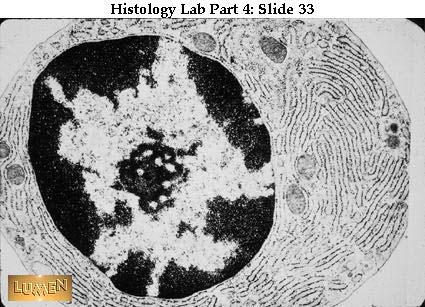
Cell OrganellesCell organelles are the little workhouses within the cell. All the functions of life take place in each individual cell. Organelles can be released by breaking the plasma membrane, through homogenization and ultracentrifuging the mixture. The organelles are of different size and density and will settle out at specific rates. Overview of organelles [list="color: rgb(255, 238, 221); font-family: Arial, Tahoma, Helvetica, FreeSans, sans-serif; font-size: 13.524px; text-align: right; background-color: rgb(68, 21, 0);"] [*] The nucleus is in the center of most cells. Some cells contain multiple nuclei, such as skeletal muscle, while some do not have any, such as red blood cells. The nucleus is the largest membrane-bound organelle. Specifically, it is responsible for storing and transmitting genetic information. The nucleus is surrounded by a selective nuclear envelope. The nuclear envelope is composed of two membranes joined at regular intervals to form circular openings called nuclear pores. The pores allow RNA molecules and proteins modulating DNA expression to move through the pores and into the cytosol. The selection process is controlled by an energy-dependent process that alters the diameter of the pores in response to signals. Inside the nucleus, DNA and proteins associate to form a network of threads called chromatin. The chromatin becomes vital at the time of cell division as it becomes tightly condensed thus forming the rodlike chromosomes with the enmeshed DNA. Inside the nucleus is a filamentous region called the nucleolus. This serves as a site where the RNA and protein components of ribosomes are assembled. The nucleolus is not membrane bound, but rather just a region. [/list] [list="color: rgb(255, 238, 221); font-family: Arial, Tahoma, Helvetica, FreeSans, sans-serif; font-size: 13.524px; text-align: right; background-color: rgb(68, 21, 0);"] [*] Ribosomes are the sites where protein molecules are synthesized from amino acids. They are composed of proteins and RNA. Some ribosomes are found bound to granular endoplasmic reticulum, while others are free in the cytoplasm. The proteins synthesized on ribosomes bound to granular endoplasmic reticulum are transferred from the lumen (open space inside endoplasmic reticulum) to the golgi apparatus for secretion outside the cell or distribution to other organelles. The proteins that are synthesized of free ribosomes are released into the cytosol. [/list] ـــــــــــــــــــــــــــــــــــــــــــــــــــــــــــــــــــــــــــــــــــــــــــــــــــــــــــــــــــــــــــــــــــــــــــــــــــ [list="color: rgb(255, 238, 221); font-family: Arial, Tahoma, Helvetica, FreeSans, sans-serif; font-size: 13.524px; text-align: right; background-color: rgb(68, 21, 0);"] [*] The gol gi apparatus gi apparatus is a [/list] 
gilgi appratus[list="color: rgb(255, 238, 221); font-family: Arial, Tahoma, Helvetica, FreeSans, sans-serif; font-size: 13.524px; text-align: right; background-color: rgb(68, 21, 0);"] [*] membranous sac that serves to modify and sort proteins into secretory/transport vesicles. The vesicles are then delivered to other cell organelles and the plasma membrane. Most cells have at least one golgi apparatus, although some may have multiple. The apparatus is usually located near the nucleus. [/list] ـــــــــــــــــــــــــــــــــــــــــــــــــــــــــــــــــــــــــــــــــــــــــــــــــــــــــــــــــــــــــــــــــــ
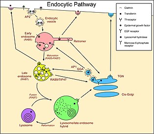

 [list="color: rgb(255, 238, 221); font-family: Arial, Tahoma, Helvetica, FreeSans, sans-serif; font-size: 13.524px; text-align: right; background-color: rgb(68, 21, 0);"] [*] Endosomes are membrane-bound tubular and vesicular structures located between the plasma membrane and the golgi apparatus. They serve to sort and direct vesicular traffic by pinching off vesicles or fusing with them. [/list] ــــــــــــــــــــــــــــــــــــــــــــــــــــــــــــــــــــــــــــــــــــــــــــــــــــــــــــــــــــــــــــــــــ

 [list="color: rgb(255, 238, 221); font-family: Arial, Tahoma, Helvetica, FreeSans, sans-serif; font-size: 13.524px; text-align: right; background-color: rgb(68, 21, 0);"] [*] Lysosomes are bound by a single membrane and contain highly acidic fluid. The fluid acts as digesting enzymes for breaking down bacteria and cell debris. They play an important from in the cells of the immune system. [/list] ــــــــــــــــــــــــــــــــــــــــــــــــــــــــــــــــــــــــــــــــــــــــــــــــــــــــــــــــــــــــــــــ [list="color: rgb(255, 238, 221); font-family: Arial, Tahoma, Helvetica, FreeSans, sans-serif; font-size: 13.524px; text-align: right; background-color: rgb(68, 21, 0);"] [*] Peroxisomes are also bound by a single membrane. They consume oxygen and work to drive reactions that remove hydrogen from various molecules in the form of hydrogen peroxide. They are important in maintaining the chemical balances within the cell. [/list] [list="color: rgb(255, 238, 221); font-family: Arial, Tahoma, Helvetica, FreeSans, sans-serif; font-size: 13.524px; text-align: right; background-color: rgb(68, 21, 0);"]
Microfilaments are the thinnest and most abundant of the cytoskeleton proteins. They are composed of actin, a contractile protein, and can be assembled and disassembled quickly according to the needs of the cell or organelle structure.
[/list] [list="color: rgb(255, 238, 221); font-family: Arial, Tahoma, Helvetica, FreeSans, sans-serif; font-size: 13.524px; text-align: right; background-color: rgb(68, 21, 0);"]
Intermediate filaments are slightly larger in diameter and are found most extensively in regions of cells that are going to be subjected to stress. Desmosomes in the skin will contain filaments. Once these filaments are assembled they are not capable of rapid disassembly.
[/list] [list="color: rgb(255, 238, 221); font-family: Arial, Tahoma, Helvetica, FreeSans, sans-serif; font-size: 13.524px; text-align: right; background-color: rgb(68, 21, 0);"]
Microtubules are hollow tubes composed of a protein called tubulin. They are the thickest and most rigid of the filaments. Microtubules are present in the axons and long dendrite projections of nerve cells. They are capable of rapid assembly and disassembly according to need. Microtubules are structured around a cell region called the centrosome, which surrounds two centrioles composed of 9 sets of fused microtubules. These are important in cell division when the centrosome generates the microtubluar spindle fibers necessary for chromosome separation.
[/list] ـــــــــــــــــــــــــــــــــــــــــــــــــــــــــــــــــــــــــــــــــــــــــــــــــــــــــــــــــــــــــــــــــــــــــ [list="color: rgb(255, 238, 221); font-family: Arial, Tahoma, Helvetica, FreeSans, sans-serif; font-size: 13.524px; text-align: right; background-color: rgb(68, 21, 0);"] [*] The endoplasmic reticulum (ER) is collectively a network of membranes enclosing a singular continuous space. As mentioned earlier, granular endoplasmic reticulum is associated with ribosomes (giving the exterior surface a rough, or granular appearance). Sometimes granular endoplasmic reticulum is referred to as rough ER. The granular ER is involved in packaging proteins for the golgi apparatus. The agranular, or smooth, ER lacks ribosomes and is the site of lipid synthesis. In addition, the agranular ER stores and releases calcium ions Ca 2+ . [/list] ــــــــــــــــــــــــــــــــــــــــــــــــــــــــــــــــــــــــــــــــــــــــــــــــــــــــــــــــــــــ Mitochondria [list="color: rgb(255, 238, 221); font-family: Arial, Tahoma, Helvetica, FreeSans, sans-serif; font-size: 13.524px; text-align: right; background-color: rgb(68, 21, 0);"] [*] Mitochondria are some of the most important structures in the human body. They are they site of various chemical processes involved in the synthesis of energy packets called ATP (adenosine triphosphate). Each mitochondrion is surrounded by two membranes. The outer membrane is smooth, while the inner one is folded into tubule structures called cristae. Mitochondria are unique in that they contain small amounts of DNA containing the genes for the synthesis of some mitochondrial proteins. The DNA is inherited solely from the mother. Cells with greater activity have more mitochondria, while those that are less active have less need for energy producing mitochondria. [/list] Mitochondria are found exclusively in eukaryotic cells. These organelles are often called the "power plants" of the cell because their main job is to make energy (ATP). Mitochondria are highly unusual--they contain their own genetic material and protein-making machinery enwrapped in a double membrane ــــــــــــــــــــــــــــــــــــــــــــــــــــــــــــــــــــــــــــــــــــــــــــــــــــــــــــــــ , cilia are hair-like motile extensions on the surface of some epithelial cells. They have a central core of 9 sets of fused microtubules. In association with a contractile protein, these microtubules produce movement in cilia. Ciliar movements propel the luminal contents of hollow organs lined with ciliated epithelium
- Flagella & cilia - hair-like projections from some human cells
- cilia are relatively short & numerous (e.g., those lining trachea)
- a flagellum is relatively long and there's typically just one
ـــــــــــــــــــــــــــــــــــــــــــــــــــــــــــــــــــــــــــــــــــــــــــــــــــــــــــــــ  The DNA stored in the nucleus of a single human cell spans over six feet in length if stretched from end to end. Made up of four chemical building blocks called A, C, T and G, for short, DNA contains the instructions for making all living things. The building blocks link to form the molecule's famous "double helix" structure, which allows genetic information to be copied and passed down from one generation to the next. Occasionally exposure to toxins or malfunction of cellular processes, among other things, does cause copying mistakes. Such changes over long time periods provide opportunities for organisms to adapt to new surroundings--or, cause them to die out. Discrete segments of DNA, called genes, encode the instructions for making proteins. Work horses of the cell, proteins serve as structural material, hormones, enzymes and neurotransmitters as well as play many other roles  Tucked away inside the DNA of all of your genes are the instructions for how to construct a unique individual. Our genetic identity is "coded" in the sense that four building blocks, called nucleotides, string together to spell out a biochemical message—the manufacturing instructions for a protein. DNA's four nucleotides, abbreviated A, T, G, and C, can only match up in specific pairs: A links to T and G links to C. An intermediate in this process, called mRNA (messenger ribonucleic acid), is made from the DNA template and serves as a link to molecular machines called ribosomes. Inside every cell, ribosomes read mRNA sequences and hook together protein building blocks called amino acids in the order specified by the code: Groups of three nucleotides in mRNA code for each of 20 amino acids. Connector molecules called tRNA (transfer RNA) aid in this process. Ultimately, the string of amino acids folds upon itself, adopting the unique shape that is the signature of that particular protein.    Transcription - DNA is used to produce mRNA [*] sequence of amino acids in a protein is determined by sequence of codons (mRNA). Codons are 'read' by  anticodons of tRNAs & tRNAs then 'deliver' their amino acid. anticodons of tRNAs & tRNAs then 'deliver' their amino acid. [*] Amino acids are linked together by peptide bonds (see diagram to the right) [*] As mRNA slides through ribosome, codons are exposed in sequence & appropriate amino acids are delivered by tRNAs. The protein (or polypeptide) thus grows in length as more amino acids are delivered. [*] The polypeptide chain then 'folds' in various ways to form a complex three-dimensional protein molecule that will serve either as a structural protein or an enzyme ـــــــــــــــــــــــــــــــــــــــــــــــــــــــــــــــــــــــــــــــــــــــــــــــــــــــــــــــــــــــــــــ Active Transport: The Sodium-Potassium Pump

[*]Endo- & exocytosis - moving material into (endo-) or out of (exo-) cell in bulk form
[*] ــــــــــــــــــــــــــــــــــــــــــــــــــــــــــــــــــــــــــــــــــــــــــــــــــــــــــــــــــــــــــ

Mitochondrial electron transport chain. Notice that the 'chain' of reactions that occurs with the conversion of NADH to NAD+ result in the transport of three pairs of hydrogens (2H+) (that will then result in the production of 3ATP), whereas the reactions occuring after the conversion of FADH2 to FAD result in the transport of two 2H+

The cellular metabolism of substrates such as glucose and fatty acids (green arrows in the figure) generates hydrogens and, specifically, hydrogen carriers — NADH and FADH2. NADH and FADH2 donate electrons to the electron-transport chain that consists of proteins located in the mitochondrial inner membrane. Electrons are ultimately transported to molecular oxygen that is reduced to water in the last step of the electron-transport chain. As electrons are transferred along the electron-transport chain, the energy released is used to pump protons (H+) from the mitochondrial matrix into the mitochondrial intermembrane space. The energy produced drives the synthesis of ATP from ADP and inorganic phosphate (Pi) by ATP synthase. ATP is then made available to the cell for various processes (e.g., active transport)

ATP synthase. The proton channel and rotating stalk are shown in blue
ـــــــــــــــــــــــــــــــــــــــــــــــــــــــــــــــــــــــــــــــــــــــــــــــــــــــــــــــــــــــــــــــــــــــــــ


|
| | | | ابراهيم الشنطي
Admin
عدد المساهمات : 70301
تاريخ التسجيل : 28/01/2013
العمر : 77
الموقع : الاردن
| | موضوع: رد: صور طبية هيستولوجى - Histology الإثنين 19 نوفمبر 2018, 12:15 am | |
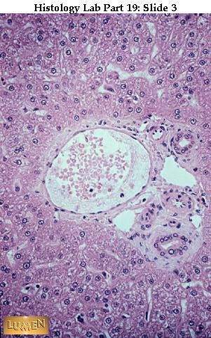
| : Liver, Salivaries, and Pancreas
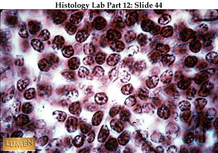
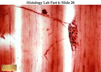
Typical liver lobule, with cords of parenchymal cells radiating from central vein. (H&E stain).
Liver lobule outlined by blue connective tissue septa. Central vein is in the center. portal canals lie out at the "corners", in the connective tissue. (Mallory stain)
Portal canal (triad) as seen in liver of most mammals, including humans. The largest vessel is a branch of the portal at upper left and right of portal area are two branches of bile duct with simple cuboidal lining; between them lies a branch of the hepatic artery with pink tunica media (smooth muscle). The irregular-shaped empty spaces with endothelial linings are lymphatics.
Another portal area with branches of the portal vein (largest), the hepatic artery (to right), and bile duct (above, center - with cuboidal epithelium)
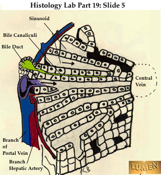
Diagram of arrangement of lobule. A portal triad is at the left. Blood in the branches of the hepatic artery and portal vein enters the sinusoids, between the cords of liver cells, and courses toward the central vein, which is a tributary of the hepatic vein. Bile flows in the opposite direction, from the center out, toward the tributaries of the bile duct. Bile canaliculi are tiny channels which exist between the cell surfaces of neighboring hepatic parenchymal cells. "Parenchyma" means the working cells of an organ - in this case, the hepatic cord cells (hepatocytes). They are supported by a delicate connective tissue stroma.
 Detail of hepatic cords with sinusoids coursing between them. The lining endothelial cells have pulled away from the cord cells somewhat, leaving an enlarged space of Disse, which ordinarily could be seen only at the EM level. The nuclei associated with the sinusoids here may belong either to endothelial cells or to macrophages (the Kupffer cells). The blue at the top of the field is connective tissue of Glisson's capsule; this is out at the edge of the liver.
Diagram of EM of liver cell and its contacts. Notice that some surfaces lie next to a space of Disse and a sinusoid (as at the top), while others are next to another hepatocyte and include a small bile channel (see left and right sides). The bile canaliculi have no other wall than the cell surfaces of the of the hepatocytes.
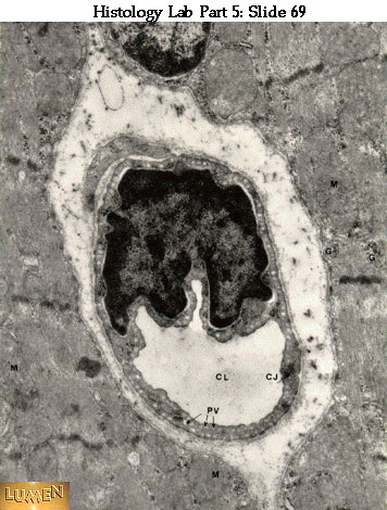
EM of bile canaliculus -- the empty spot between adjacent hepatocytes. Essentially, it is a widening of the intercellular space. Notice the typical organellar composition of the adjoining hepatocytes cytoplasm.
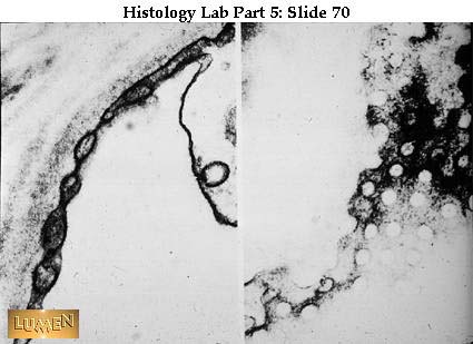
Detail of silvered bile canaliculi. Notice the little hollow channels lying between adjacent hepatocytes. Where the channels are cut in cross- section you'll see tiny circles. Clearly, these canaliculi form an all- pervasive network throughout the liver. Between the double rows of hepatocytes lie sinusoids with black red blood cells in them.
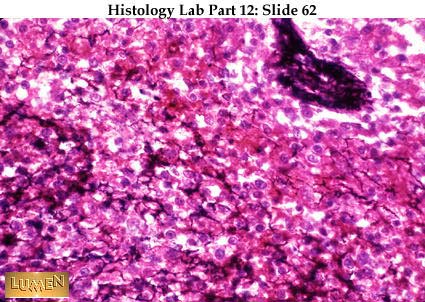
Detail of hepatic cords stained in ordinary H&E, showing a cross-cut bile canaliculus between two cells near the upper right corner of the field. (Look for a tiny oval opening in the horizontal line between cells near the top edge of the field; a dark, tight junction seals the oval at each end.) Large intercellular spaces elsewhere in the field are sinusoids.
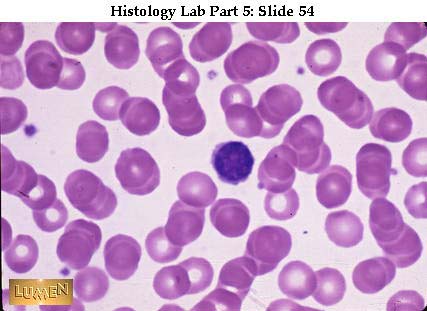
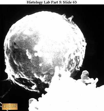
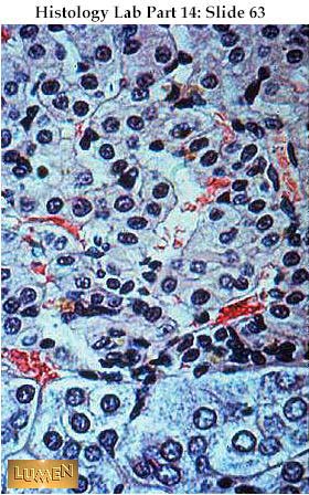
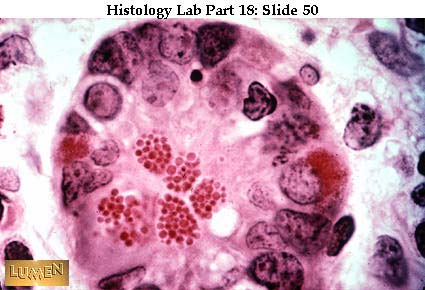
Kupffer cells lying in sinusoids and loaded with phagocytized carbon particles.
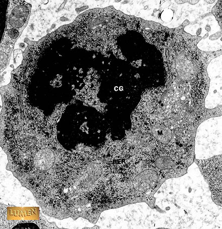
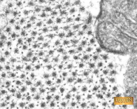
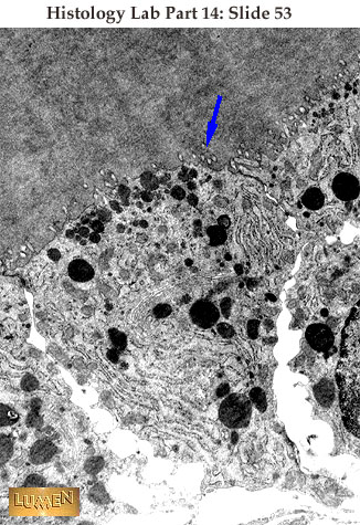
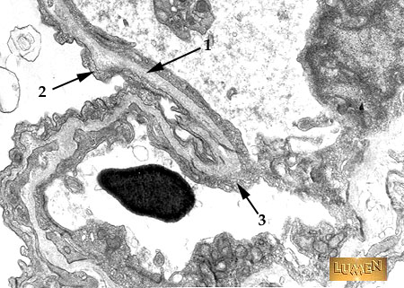
EM of liver cell (hepatocyte), showing aggregates of glycogen (1). Compare their size with the ribosomes lining the endoplasmic reticulum (2).
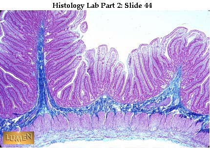
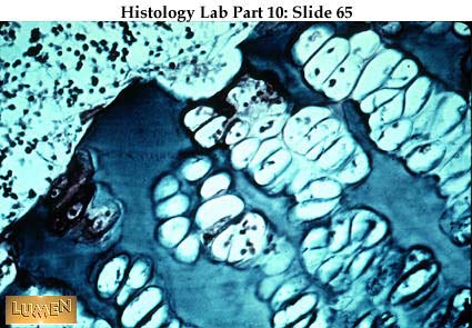
GallbladderWall of gall bladder, showing high, branching mucosal folds. These are not villi. The rest of the wall contains connective tissue and thin strands of smooth muscle.
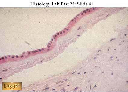
Detail of mucosal fold with very clear simple columnar epithelium - no goblet cells! While the epithelial cells do tend to have some microvilli on their apical border, these do not form the concentrated brush (or striated) border that is typical of cells lining the intestinal villi.
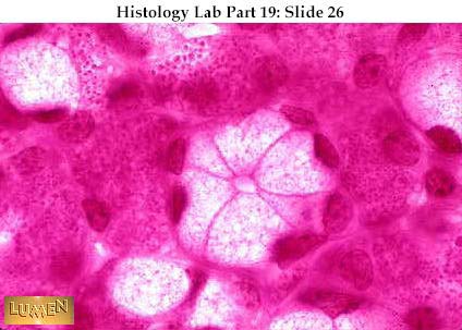
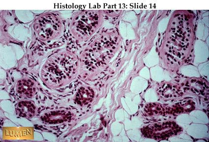
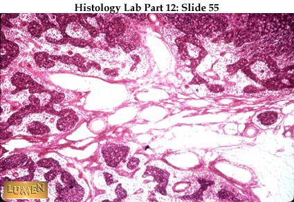
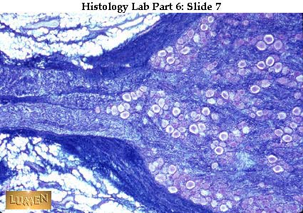
Salivary Glands Low power view to show the typical structure of a compound tubulo- alveolar gland. The pink, solid-looking masses are lobules composed of secretory alveoli (acini). Separating the lobules from each other are pale connective tissue septa. Running within the septa (as best seen in the upper left corner of this picture) are darkly staining interlobular ducts, which can be seen here and there invading the lobules as intralobular ducts. As these smaller ducts continue to branch within the substance of the lobule, they ultimately end in secretory units, the alveoli (or acini).
 Interlobular area of gland, showing several structures lying in the connective tissue. Both a longitudinal and cross cut of an interlobular duct are toward the bottom of the field, with their typical, two-layered stratified cuboidal epithelial lining. Above are several blood vessels filled with dark pink-staining red blood cells. In the upper center is quite a large parasympathetic ganglion packed with neurons. Lobules of glandular tissue are at the periphery of the field.
Mixed salivary gland, showing branching intralobular ducts.
Diagram of secretory units at the end of a small duct of salivary gland. Mucous and serous units are the things to notice here plus a serous demilune capping the mucous tubule at the right. ("Demilune" means "half moon.") Compare the position of the nuclei within serous vs. mucous cells.
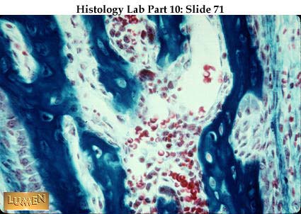
Secretory acini of mixed salivary gland (such as the submandibular gland). (The parotid gland is wholly serous.) In this stain the serous units are dark, the mucous ones light. The bright pink line to the right is a thin connective tissue interlobular septum.
In the middle is a dark, serous demilune capping a mucous acinus.
Mixed salivary gland stained with Mallory to show the bright blue areolar connective tissue stroma that supports the acini and carries capillaries and nerves. The finest fibers would be reticular. The acinar cells, both mucous and serous, constitute the parenchyma of the gland.
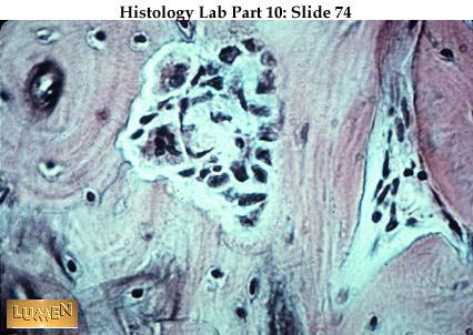
Special fixation to show zymogen granules in the serous acini; these cells are producing protein secretions (enzymes, etc.). Mucous acini are white. A pale pink duct (intralobular) is at the left.
High magnification of mucous acinus with small lumen in the center. In the serous acini occupying most of the rest of the field, red zymogen granules are distinct.
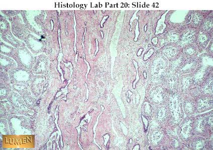
PancreasPanoramic view of pancreas showing the same kind of lobular structure seen in the salivary gland. Note interlobular vessels in the connective tissue septa.
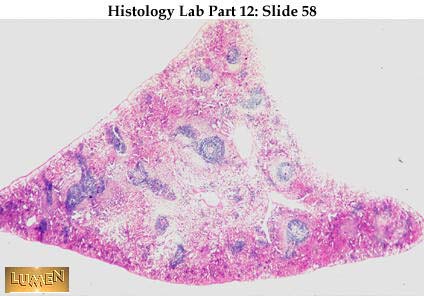
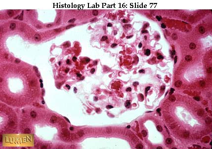
Another view of pancreas, with thin connective tissue septa dividing the parenchyma into lobules of secretory acini. Intralobular ducts are present but are not as clearly defined as in the salivary glands. The pancreas is essentially a serous gland. It is distinguishable from the parotid gland by the presence of scattered, pale islets of Langerhans, which constitute the endocrine portion of this organ.
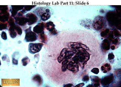
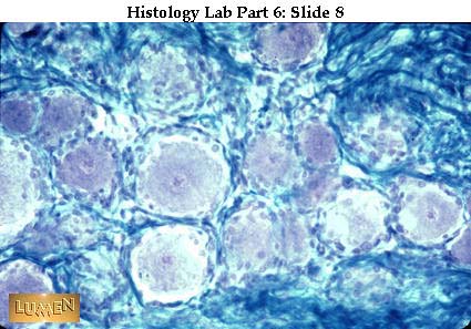
Higher magnification of a pale islet of Langerhans surrounded by secretory acini. Most of the islet cells are beta cells, which secrete insulin directly into the blood stream.
Very high power view of pancreatic acinar cells with some pale centroacinar cells in the middle of the acinus. The centroacinar cells represent the very end of the duct system, projecting into the lumen of the acinus. Only the pancreas has such an arrangement. Notice that the zymogen granules of the acinar cells lie at the apical ends; nuclei are toward the base. The granules are precursors of pancreatic digestive enzymes. These acinar cells, like the serous acinar cells of the salivary gland, are structurally like Paneth cells of the small intestines and chief cells of the stomach; all of them secrete enzymes.
EM detail of cytoplasm of pancreatic acinar cell loaded with lamellae of rough endoplasmic reticulum, typical of a protein-secreting cell. The cell nucleus is at upper left. |
| | | | ابراهيم الشنطي
Admin
عدد المساهمات : 70301
تاريخ التسجيل : 28/01/2013
العمر : 77
الموقع : الاردن
| | موضوع: رد: صور طبية هيستولوجى - Histology الإثنين 19 نوفمبر 2018, 12:16 am | |
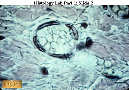
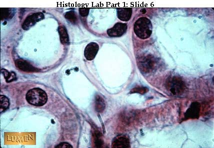
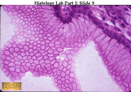
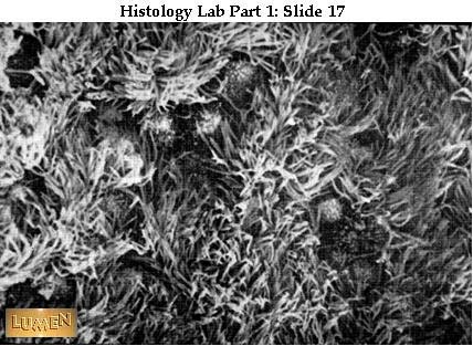
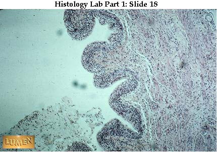
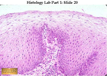
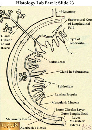
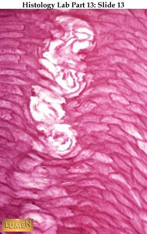
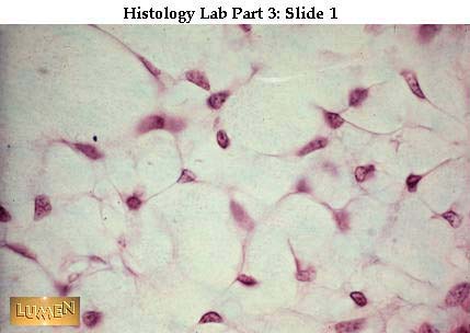
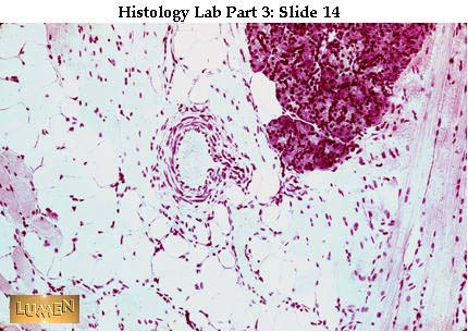
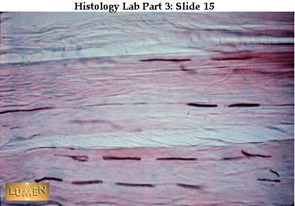
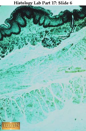
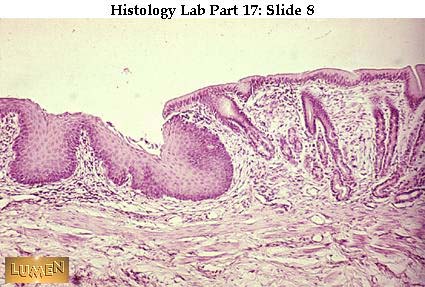
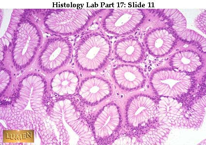
| Epithelium and Simple Glands ــــــــــــــــــــــــــــــــــــــــــــــــــــــــــــــــــــــــــ Mesothelium seen as if looking down on a surface view to see "pavement" effect of the lining cells. Silver stains the intercellular cement dark between adjacent cells. Notice how corrugated the cell membranes are. Mesothelium = the simple squamous epithelium lining body cavities and mesenteries. ـــــــــــــــــــــــــــــــــــــــــــــــــــــــــــــــــــــــــــــــــــــــــــــــــــــــــــــــــــــــــــــــــــــــــــــــــــــــــــــــــــ High power view of endothelial cells lining a small blood vessel cut in cross-section. (You see just the nuclei - the cytoplasm between them is extremely flat.) Endothelium = the simple squamous epithelium lining blood vessels. ـــــــــــــــــــــــــــــــــــــــــــــــــــــــــــــــــــــــــــــــــــــــــــــــــــــــــــــــــــــــــــــــــــــــــــــــــــــــــــــــــ Low power view of larger vessels, showing endothelial nuclei lining the lumen. The yellowish cells filling each vessel's lumen are blood cells. ـــــــــــــــــــــــــــــــــــــــــــــــــــــــــــــــــــــــــــــــــــــــــــــــــــــــــــــــــــــــــــــــــــــــــــــــــــــــــــــــ Simple cuboidal epithelium lining a tubule (longitudinal cut). Some of the cell boundaries between "blocks" or "cubes" here are quite distinct. ــــــــــــــــــــــــــــــــــــــــــــــــــــــــــــــــــــــــــــــــــــــــــــــــــــــــــــــــــــــــــــــــــــــــــــــــــــــــــــ Simple cuboidal epithelium in Mallory stain (longitudinal cut). Note the dark chromatin clumps in the nuclei. Underneath the epithelium lies a small blood vessel filled with orange-colored blood cells. ــــــــــــــــــــــــــــــــــــــــــــــــــــــــــــــــــــــــــــــــــــــــــــــــــــــــــــــــــــــــــــــــــــــــــــــــــــــــــــــــــ Cross-section of tubules. The smaller ones clustered in the center and upper left are lined by simple squamous epithelium. The larger pink tubules have simple cuboidal epithelium. ـــــــــــــــــــــــــــــــــــــــــــــــــــــــــــــــــــــــــــــــــــــــــــــــــــــــــــــــــــــــــــــــــــــــــــــــــــــــــــــــــــــــ A tubule stained to show the pink basement membrane underlying the base of the simple cuboidal epithelium. Stained with periodic acid Schiff reagent (PAS), which stains mucopolysaccharides. ـــــــــــــــــــــــــــــــــــــــــــــــــــــــــــــــــــــــــــــــــــــــــــــــــــــــــــــــــــــــــــــــــــــــــــــــــــــــــــــــــــ Simple columnar epithelium with very regular line-up of nuclei. ـــــــــــــــــــــــــــــــــــــــــــــــــــــــــــــــــــــــــــــــــــــــــــــــــــــــــــــــــــــــــــــــــــــــــــــــــــــــــــــــــــــــــــــــ Simple columnar cells cut tangentially to show how they form a very regular "pavement" when viewed from the surface. The cells are like tall blocks arranged very closely to each other with a small amount of tissue fluid in between. ــــــــــــــــــــــــــــــــــــــــــــــــــــــــــــــــــــــــــــــــــــــــــــــــــــــــــــــــــــــــــــــــــــــــــــــــــــــــــــ Detail of simple columnar epithelium with striated border (microvilli). Notice that the border is quite thin and the striations close together, looking like very regular, closely set brush bristles. ــــــــــــــــــــــــــــــــــــــــــــــــــــــــــــــــــــــــــــــــــــــــــــــــــــــــــــــــــــــــــــــــــــــــــــــــــــــــــــــــــــــــــ EM of cells with striated border. Notice the evenness and regularity of the microvilli. This is an adaptation of the cell surface for absorption. Notice also the corrugation of the cell boundaries as they fit next to each other. 1= nucleus; 2=brush border (microvilli); 3=lymphocyte. ــــــــــــــــــــــــــــــــــــــــــــــــــــــــــــــــــــــــــــــــــــــــــــــــــــــــــــــــــــــــــــــــــــــــــــــــــــــــــــــــ Detail of simple columnar epithelium with a goblet cell secreting mucus. The thin, clearly defined band along the top epithelial surface is the striated border, though the individual striations (or microvilli) are not visible at this magnification. The lower edge of the striated border is the location of the terminal web; the dots along the line of the web, seen in between the individual epithelial cells, are the so-called terminal bars, which are found in EM to consist of various cell junctions. ـــــــــــــــــــــــــــــــــــــــــــــــــــــــــــــــــــــــــــــــــــــــــــــــــــــــــــــــــــــــــــــــــــــــــــــــــــــــــــــــــــ EM of apical (top) surface of two epithelial cells whose cell membranes lie next to each other. The microvilli (1) of the striated border are very straight and regimented in appearance. Microfilaments within them can be seen extending down into the terminal web (2), which is an aggregate of fine filaments lying in the cell cytoplasm. Several junctional complexes are seen including tight junction (zonula occludens =3); intermediate junction (zonula adherens =4); and desmosome (macula adherens =5). ــــــــــــــــــــــــــــــــــــــــــــــــــــــــــــــــــــــــــــــــــــــــــــــــــــــــــــــــــــــــــــــــــــــــــــــــــــــــــــــــــــــــ  Four rows of simple columnar epithelium facing each other in pairs (left and right) across a narrow lumen or channel that lies in the middle of each pair. (This is a Mallory Trichrome stain.) The goblet cells are filled with blue mucoid secretion which is being poured into the narrow lumens. Notice that in all four rows of epithelium there is a narrow band of striated border next to the lumen; the dark purple line at the base of the border is the terminal web. Look at the right hand rows of epithelial cells and notice the dark dots all along the terminal web lines; these dots represent the junctional complexes between cells. The central cavity in the picture is a blood vessel with endothelium, surrounded by a very cellular connective tissue. Separating this connective tissue from the epithelium is a thin blue layer of connective tissue fibers. ــــــــــــــــــــــــــــــــــــــــــــــــــــــــــــــــــــــــــــــــــــــــــــــــــــــــــــــــــــــــــــــــــــــــــــــــــــــــــــــــــــــــــــ Pseudostratified ciliated columnar epithelium from the trachea. Nuclei are at different levels. All cells touch the basement membrane, but only the taller cells reach the lumen. The cilia are longer and less regular than the microvilli of a striated border. ـــــــــــــــــــــــــــــــــــــــــــــــــــــــــــــــــــــــــــــــــــــــــــــــــــــــــــــــــــــــــــــــــــــــــــــــــــــــــــــــــــ Pseudostratified ciliated columnar epithelium with pale goblet cells. The different levels of nuclei are clearer here. Again, notice the wavy-looking cilia. ـــــــــــــــــــــــــــــــــــــــــــــــــــــــــــــــــــــــــــــــــــــــــــــــــــــــــــــــــــــــــــــــــــــــــــــــــــــــــــــــــــــــــــ Surface view of cilia with scanning EM scope. Notice how "ragged" the surface seems -- cilia were caught as they moved. ـــــــــــــــــــــــــــــــــــــــــــــــــــــــــــــــــــــــــــــــــــــــــــــــــــــــــــــــــــــــــــــــــــــــــــــــــــــــــــــــــــــ Transitional epithelium of the urinary bladder, low power view. It is a stratified epithelium with several layers of cells. ــــــــــــــــــــــــــــــــــــــــــــــــــــــــــــــــــــــــــــــــــــــــــــــــــــــــــــــــــــــــــــــــــــــــــــــــــــــــــــــــــــــــــــــــ Transitional epithelium, high power. Notice many layers of cells -- and the typically puffy surface cells. The bladder is contracted so the epithelium is thick. If the bladder were stretched, the epithelium would be thinner. ــــــــــــــــــــــــــــــــــــــــــــــــــــــــــــــــــــــــــــــــــــــــــــــــــــــــــــــــــــــــــــــــــــــــــــــــــــــــــــــــــــ Stratified squamous non-cornified epithelium -- medium power. This is from the esophagus, so the surface is moist and living. Surface cells are squamous and still nucleated. Basal layer is very distinct; compare this with the less distinct basal layer of the preceding slide of transitional epithelium. ــــــــــــــــــــــــــــــــــــــــــــــــــــــــــــــــــــــــــــــــــــــــــــــــــــــــــــــــــــــــــــــــــــــــــــــــــــــــــــــــــــــــــــــ Stratified squamous epithelium with beginning surface cornification. This section is from thin skin, which has a dry surface covered with dead cells. Notice how flat the surface cells are and how dark and pyknotic their nuclei have become. Again, notice the distinct row of basal cells. ـــــــــــــــــــــــــــــــــــــــــــــــــــــــــــــــــــــــــــــــــــــــــــــــــــــــــــــــــــــــــــــــــــــــــــــــــــــــــــــــــــــــــ Thickly cornified stratified squamous epithelium. The cells in the bright red layer and in the pale layers above it are completely flattened and dead, and have lost their nuclei. ـــــــــــــــــــــــــــــــــــــــــــــــــــــــــــــــــــــــــــــــــــــــــــــــــــــــــــــــــــــــــــــــــــــــــــــــــــــــــــــــــ Diagram of GI wall to show various kinds of glands -- some within the wall and some without (like the liver). These glands have ducts that empty into the lumen of the gut. In all cases, the epithelium lining the ducts and glands is continuous with the epithelium lining the lumen (cavity) of the gut. (Note: the test-tube-like glands, labeled "crypts of Lieberkuhn" here, are the same kind as the intestinal glands you saw under the microscope in the appendix in lab.) ــــــــــــــــــــــــــــــــــــــــــــــــــــــــــــــــــــــــــــــــــــــــــــــــــــــــــــــــــــــــــــــــــــــــــــــــــــــــــــــــــــــــــــ Unicellular gland - a goblet cell mucus-secreting. (H & E stain.) ــــــــــــــــــــــــــــــــــــــــــــــــــــــــــــــــــــــــــــــــــــــــــــــــــــــــــــــــــــــــــــــــــــــــــــــــــــــــــــــــــــــــ Goblet cells (blue) scattered throughout simple columnar epithelial lining (special quad stain). ــــــــــــــــــــــــــــــــــــــــــــــــــــــــــــــــــــــــــــــــــــــــــــــــــــــــــــــــــــــــــــــــــــــــــــــــــــــــــــــــــــــــــــــــــ Simple tubular glands of gut wall seen in low power. These glands are lined with epithelium throughout their whole extent. ـــــــــــــــــــــــــــــــــــــــــــــــــــــــــــــــــــــــــــــــــــــــــــــــــــــــــــــــــــــــــــــــــــــــــــــــــــــــــــــــــــــــــ Detail of such a gland. Goblet cells are purple here. -(H & E) ـــــــــــــــــــــــــــــــــــــــــــــــــــــــــــــــــــــــــــــــــــــــــــــــــــــــــــــــــــــــــــــــــــــــــــــــــــــــــــــــــــــــــــــــــــ Low power of wall of esophagus showing duct at right, leading down to simple tubulo-alveolar gland with coiled secretory portions. ــــــــــــــــــــــــــــــــــــــــــــــــــــــــــــــــــــــــــــــــــــــــــــــــــــــــــــــــــــــــــــــــــــــــــــــــــــــــــ Drawings of compound tubulo-alveolar glands -- showing the branching of their duct system -- and a few secretory end-pieces (alveoli). Ducts and alveoli are lined with epithelium. ــــــــــــــــــــــــــــــــــــــــــــــــــــــــــــــــــــــــــــــــــــــــــــــــــــــــــــــــــــــــــــــــــــــــــــــــــــــــــــــــــــــــــــــــ High power of typical mucous (pale) and serous (darker pink) secretory cells. Notice that the nuclei of mucous cells are dark and flattened at the base of the cells, while the nuclei of serous cells are round and more centrally located at their cells. Mucous secretion is relatively thick and viscous; serous secretion is watery. |
| | | | ابراهيم الشنطي
Admin
عدد المساهمات : 70301
تاريخ التسجيل : 28/01/2013
العمر : 77
الموقع : الاردن
| | موضوع: رد: صور طبية هيستولوجى - Histology الإثنين 19 نوفمبر 2018, 12:16 am | |
| [size=32]Slides of Skin and Tongue[/size]
Thick skin, to be compared with the next slide (thin skin) for thickness of epidermis. Notice also in each slide a duct of sweat gland going down through the dermis.
Thin skin at the same magnification as previous slide. The sweat duct in this case is clearly ending at its coiled secretory portion. Hypodermis in both slides is just beginning at the bottom of the pictures, where you begin to see some fat cells.
Thick skin showing epithelial detail. Cornified (keratinized) stratified squamous epithelium makes up the epidermis. The stratum granulosum is very dark; the stratum lucidum is bright red. The stratum corneum is thick, and very pale.
Detail of epithelium of thick skin from the superficial to deep:
- pale stratum corneum
- bright red stratum lucidum
- purple stratum granulosum
- stratum spinosum
Note the basophilic, keratohyaline granules in the stratum granulosum; their presence indicates that the cells in this layer are beginning to die
Stratum spinosum showing "prickle" appearance of cell contacts. (The cells of this layer are often called "prickle cells".) The cross-lines were once thought to be intercellular bridges.
EM detail of "prickle cell" contact, showing the presence of many desmosomes between cells (arrows). The little fibrillar strands lying in the cytoplasm and inserting on the desmosomes are tonofilaments (F). The cell membranes of the two cells are repeatedly interdigitated, giving the appearance of "intercellular bridges" in light microscopy.
Detail of epithelium of thin skin, showing melanin in the basal layer. The pigment is produced by stellate shaped melanocytes of the dermal layer and then deposited in the basal cells of the epidermis. Melanocytes are of neural crest origin and have to be specifically stained in order to be seen.
Thin skin showing epithelial detail. This is also cornified stratified squamous, but thinner. The granulosum is only about one cell layer thick and is the darkest layer here. There is no stratum lucidum.
Detail of thin skin epithelium. A single layer of dark purple granulosum lies under the rather shredded stratum corneum. The stratum corneum typically sloughs off with wear. Stratum spinosum is at the bottom of the picture.
The next few pictures are various views of sweat glands. This one shows a long straight sweat duct heading down through the dermis. Its epithelial lining is continuous with the basal cells of the epidermal epithelium.
 The secretory portions of several sweat glands lie in clusters among the fat cells of the hypodermis, low in the picture. (A bedraggled, shrunken Pacinian corpuscle is in the same area, just left of center.) Sweat glands are simple coiled tubules, so we see many cross-cuts of the coil in a section like this. These are the true (eccrine) sweat g lands of skin and therefore secrete their watery (serous) fluid by merocrine secretion. (Some of the very large sweat glands of the axillary and pubic regions are thought to use apocrine secretion, pinching off small bits of cell cytoplasm or membrane along with the secretory product.) While looking at this picture, note the very dense, irregular connective tissue of the dermis. Right under the epidermis notice a brighter, clearer, quite narrow, pink layer of dermis; this is the papillary layer of the dermis and it forms the dermal papillae which extend up into irregularities of the basal layer of the epidermis, bringing capillary loops and nerve endings closer to the epithelial cells an d to the surface. The bulk of the dermis, below this thinner layer, is the reticular layer; it is more heavily fibrous, with many elastic fibers running among the more numerous collagen fibers. The hypodermis begins with the fat cells.
 Detail of sweat duct originating (at lower left) from the basal layer of the epidermal epithelium.
Detail of sweat duct coiling throughout the stratum corneum to the surface of the skin. Since cells are dead here, there is no longer a living duct lining; just the tunneling of its lumen remains.
Detail of sweat gland. The darker circles in the lower part of the field are ducts; the lighter cross-cuts above are the secretory portions.
Three-dimensional drawing of dark, spidery myoepithelial cells surrounding sweat gland tubule. By contracting, they help to squeeze out the secretion.
Detail of myoepithelial cell processes as seen in H&E section. Look at the large tubule on the left for pink "hoops" that seem to be extending from the basement membrane of the upper row of epithelium toward the secretory cell nuclei which lie near the lumen. These "hoops" are the cytoplasmic extensions of the stellate myoepithelial cells. Thes e cells lie within the basal lamina of the tubule.
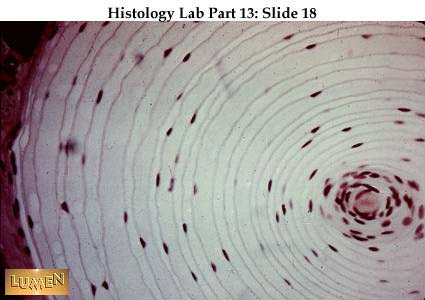
Detail of Pacinian corpuscle. Note the onion-like layers of the specialized capsule. The nerve ending itself is buried in the center (which looks pink here). The cell body for this dendritic ending lies in a spinal ganglion related to this particular dermatome of skin.
Whole mount of a Pacinian corpuscle, to give a more three-dimensional view. The dendritic ending is the darker "rod" in the middle.
Section of thick skin, as of finger tip. Projections of pale-staining dermal connective tissue push up into the bottom layers of the darker staining epithelium, carrying capillaries and nerve endings with them. In the projection on the farthest left, you can see an encapsulated Meissner's corpuscle, a sensory receptor ending for touch. Speciali zed connective tissue cell nuclei can be seen running in a circular direction around the corpuscle. The cell body for this ending lies in a spinal ganglion related to this particular dermatome of skin. (NOTE: In the stratum granulosum of this epidermis, the cells are stained dark purple.)
 Detail of Meissner's corpuscle lying in dermal papilla. The arrows point to nuclei of the specialized connective tissue sheath that surrounds the dendritic ending that is twining around inside, among the sheath cells. Silver stain would make the ending visible. You get the feeling that this corpuscle has some substance to it, i.e. that you coul d shell it out as a more or less solid unit, from the surrounding loose areolar connective tissue.
Hair follicles of scalp, with associated pale sebaceous glands. The follicles extend down into the hypodermis, which is largely adipose tissue. Notice the arrector pili muscle running diagonally toward the upper right-hand corner of the field. Its lower end would at some point attach to the follicle sheath.
Cross-sections of many hair follicles. The yellow centers are the hairs themselves. The surrounding pink cellular sheaths are continuous with the surface epithelium of the skin. A clear pink connective tissue sheath lies outside the epithelial sheath. The fat cells of the hypodermis surround the follicles.
Overview of the scalp, again showing collagen fibers of dense irregular c.t. clearly. A sweat gland duct is extending down from the surface epithelium near the top center of the field. At the extreme right, a hair is seen extending from the top of a follicle, and pale sebaceous glands are emptying into the follicle lower down.
Detail of sebaceous gland. Cells look foamy because of loss of lipid droplets during tissue fixation. This gland exhibits holocrine secretion, in which whole cells swell up, degenerate, and are desquamated as part of the oily secretion (sebum). The secretion is emptied into the hair follicles and eventually reaches the surface of the skin.
View of tongue, showing location of papillae. Most of the surface of the human tongue is covered with filiform papillae, with some fungiform papillae interspersed. A row of circumvallate papillae lie toward the back of the tongue. The lymphoid tissue labelled is lingual tonsil.
Cut-away section of tongue to show three-dimensional view of papillae and underlying c.t. and muscle.
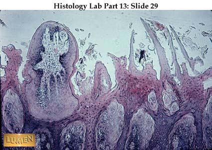
Section of surface of tongue, showing one rather tangentially cut fungiform papilla at the left and some filiform papillae with sharp, semicornified tips at the right. Cornification is less extensive in human tongue than in cats, dogs, etc.
Higher magnification of tongue surface, showing two filiform papillae. They are obviously extensions of stratified squamous epithelium.
View of foliate papillae, typical of rabbit and some other animals. These have a characteristic 3-pronged connective tissue pattern extending up into the papilla, and there are taste buds on the outside walls. Notice the bundles of skeletal muscle down below.
Detail of skeletal muscle and secretory glands of the body of the tongue. Mucous cells are to the left, with their flattened, basal nuclei, while serous cells are in the center and to the right, with their round nuclei.
Section of tongue through circumvallate papilla. Notice glands immediately below it; also the interlacing skeletal muscle strands deeper in the section.
Detail of circumvallate papilla, showing pale taste buds opening into the lumen of the furrow that surrounds the papilla.
Higher magnification of taste buds (from the foliate papillae of rabbit in this case). In the lower buds note surface pores through which salivary fluids in the lumen of the furrow reach sensory nerve endings within the taste bud capsule. The cell bodies for these dendritic endings are pseudounipolar and lie within the sensory ganglion of a cran ial nerve (such as Nerve VII). |
| | | | ابراهيم الشنطي
Admin
عدد المساهمات : 70301
تاريخ التسجيل : 28/01/2013
العمر : 77
الموقع : الاردن
| | موضوع: رد: صور طبية هيستولوجى - Histology الإثنين 19 نوفمبر 2018, 12:17 am | |
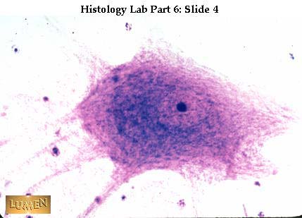
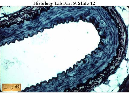
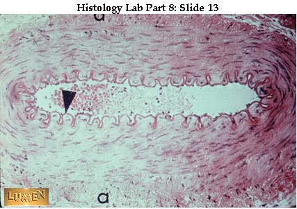
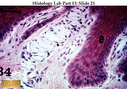
| Male Reproductive Tract  [size=13]Low power view of the testis at the mediastinal region where the duct system leaves the organ. Seminiferous tubules of the testis. [/size]
- a = spermatogonia
- b = spermatocytes (probably primary because secondary spermatocytes go through their cell division so quickly that they are seldom seen in sections). The cells are largest at this stage.
- e = spermatids
- f = maturing sperm
- d = interstitial cells of Leydig (endocrine cells which secrete androgens).
[size] A small amount of smooth muscle around the tubule aids in moving differentiated sperm along the tubule and into the rete testis  Detail of wall of seminiferous tubule, showing stages of spermatogenesis. The large, vertically oriented nucleus at the base on the left belongs to a Sertoli cell; notice that sperm heads (somewhat out of focus) are clustered deep down near this nucleus. The horizontally flattened nuclei along the base of the tubule belong to spermatogonia, the continually multiplying, diploid germ cells. A few primary spermatocytes, with the dark, condensed chromosomes undergoing prophase of meiotic division, lie just above the spermatogonia. Above the spermatocytes are the round, relatively small, haploid spermatids, which would occupy the rest of the layers up toward the lumen.  Drawing of stages in the differentiation of sperm directly from spermatids. This process is called spermiogenesis. [/size]
- A = a still-rounded spermatid, with an acrosomal body beginning to form in the region of the Golgi apparatus. At the opposite pole of the nucleus lie the centrioles, one of which begins to spin out a long cilium (or flagellum).
- B = an elongating spermatid, with an acrosomal cap now forming over the top of the nucleus. The flagellum is longer and the centrioles are oriented perpendicular to each other. The centriole related to the flagellum is comparable to the basal body of ordinary cilia.
- C = further development of the acrosomal cap and beginning pinching off of excess cytoplasm, thanks to the formation of a filamentous manchette, nuclear ring, and annulus.
- D = further condensing of the nuclear chromosomal material and separating off of the excess cytoplasm. Notice that the intercellular bridge connecting this spermatid to its neighbor is still intact.
- E = the cytoplasmic mitochondria have now collected along the proximal portion of the flagellum and are thus conserved (for energy purposes) when the residual cytoplasm is cast off. The flagellum has meanwhile developed a complex fibrous sheath which surrounds a central core of microtubules arranged in the nine plus two arrangement typical of cilia
[size]  Scanning EM of differentiated sperm in the top photos, and a drawing below of a spermatozoan as seen in transmission EM. In the SEM views: [/size]
- HR = head region, with acrosomal cap nearly covering the nucleus. This cap contains hyaluronidase, which will be released at the time of fertilization as an aid to breaking down membranes around the egg.
- NK = neck region, where the centrioles lie
- MP = middle piece, where the mitochondria congregate
- PP = principal piece of the flagellum
- EP = end-piece of the flagellum
[size] Additional labels of importance in the TEM drawing include: [/size]
- Ac = acrosomal cap
- N = nucleus
- Ne = extension of the nuclear envelope
- Ce = centriole
- M = mitochondria
- ODF = outer dense fibers of the flagellum
- FS = fibrous sheath of the flagellum
- F = flagellar core containing the 9 + 2 arrangement of microtubules (otherwise known as the axoneme).
- Mt = microtubules as seen in cross-section
[size] Cross-cuts of the three main parts of the flagellum are shown. Notice that a plasma membrane covers the entire sperm; in other words, this is a highly differentiated single cell. The flagellum is not motile when sperm leave the testis, but maturation takes place during the long stay in the epididymis. Actually, sperm are not fully capacitated for fertilization until they encounter the fluids of the vagina  Detail of EM of the three main segments of the flagellum. [/size]
- MS = mitochondrial sheath
- AF = axoneme, made of microtubules (9 + 2)
- OCF = outer course fibers
- LC + CF = parts of fibrous sheath
- P = plasmalemma
[size]  Another detailed view of spermatozoan, with appropriate cross-cuts. Notice again how the acrosomal cap (or enzyme-containing sac) fits over the nucleus.  Diagram of the relation between Sertoli cells and the cells undergoing spermatogenesis in the wall of the seminiferous tubule. Best seen to the left is a tall columnar Sertoli cell, with a very indented, irregular cell outline. Its nucleus lies near the base. The spermatogenic cells lie in the indentations of the Sertoli cell, apparently taking some nourishment from the columnar cell. The spaces where the spermatogonia, spermatocytes. and spermatids would lie are empty here, but you'll notice that there are connecting channels from space to space because these cells are incompletely separated during meiosis and are connected with each other by intercellular bridges, much as if in a chain of paper dolls. As the spermatids differentiate into sperm, their heads indent the Sertoli cells from the top and may push quite deeply down toward the nucleus, never breaking the Sertoli cell membrane but just being supported and protected in this way. Notice, near the base of the Sertoli cell, that the cell spreads out horizontally to meet its Sertoli cell neighbors and that bands of tight junctions develop between them (see arrows). These form the so-called "blood testis barrier", which provides an immunologically privileged environment for all the cells lying above it (toward the lumen). The diploid spermatogonia, which are genetically like most of the cells of the body, lie in the basal compartment, where they are exposed to all contents of the blood capillaries that surround the seminiferous tubule. Spermatocytes and spermatids, on the other hand, lie in the adluminal compartment, above the barrier, and are shielded from some substances in the blood. Remember, too, that these meiotically active and eventually haploid cells are genetically different from the rest of the body and would be recognized as "foreign" if not protected from blood and tissue fluids.  Wall of seminiferous tubule. Along the base can be seen small dark nuclei of spermatogonia and large, pale, ovoid or triangular nuclei of Sertoli cells, each with a prominent nucleolus. Sperm heads are imbedded in folds of Sertoli cell membrane, rather deep within the tubule wall. Sperm tails are pointing toward the tubule lumen. Primary spermatocytes have large nuclei with the condensed chromosomes in prophase, near the base of the wall. The small, round nuclei toward the lumen belong to early spermatids. Pale pink cytoplasmic cast-offs from differentiating spermatids (becoming sperm) lie next to the lumen. Look again at the two Sertoli cells farthest to the left and notice how their cytoplasm meets near the base to form the basal and adluminal compartments on either side of the junction.  Efferent ducts with their irregular epithelial border. These are the only portions of the male reproductive tract with motile cilia on the lining epithelium. Cilia help to move the sperm along toward the epididymis.  Detail of efferent duct wall, a low pseudostratified columnar epithelium with some surrounding smooth muscle. The epithelium is ciliated. Sperm lie in the lumen.  Epididymis with pseudostratified columnar epithelium with stereocilia. (Stereocilia are structurally like microvilli rather than like true cilia. They do not move.) Each cross-cut of tubule shows some surrounding smooth muscle cells. Notice how very regular this epithelium is in height, making an unusually smooth apical line near the lumen. This is characteristic of epididymis. Compare this with the "scalloped" edge of efferent ducts in the previous slides. 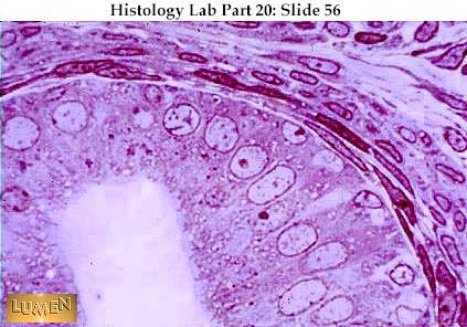 Another view of epididymal wall, showing more clearly the basal and columnar cells of the pseudostratified epithelium. The stereocilia are long and pale (practically invisible here!). Circular smooth muscle in the outer wall is evident.  Distended epididymis packed with maturing sperm. They are already mature structurally but are only now acquiring the ability to move on their own.  Low power view of spermatic cord, containing blood vessels, nerve, skeletal muscle, and a thick-walled vas deferens (ductus deferens) at the extreme upper left. 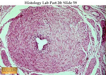 Ductus deferens with its proportionally small lumen and heavy muscular coat. The bulk of the smooth muscle is circular, but there is a thin inner longitudinal and a somewhat thicker outer longitudinal layer. 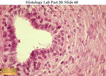 Detail of epithelial lining of the ductus deferens. It is pseudostratified columnar with non-motile stereocilia.  Prostate gland with both distended and infolded epithelial linings of the secretory portions. The saccular secretory tubules lie in a dense connective tissue framework (stained pink here). The prostate, along with the seminal vesicles and bulbo-urethral glands, contributes nourishing and lubricating fluids to the ejaculated semen.  Detail of saccular secretory tubule of prostate with a typical concretion (hardened mass) in the lumen. The concretion appears to be lamellated (in layers).  Prostate stained with Mallory to show pink/lavender strands of smooth muscle lying in the blue connective tissue framework. Notice again that the secretory tubules are wide and sacculated and that the epithelium is thrown into folds.  Penile urethra (corpus spongiosum), showing the typical dorso-ventral flattening of its lumen. The mucosal lining is highly folded. The bulk of the wall is composed of erectile tissue consisting of convoluted trabeculae surrounded by irregular vascular spaces (mainly venous sinuses) which are lined with endothelium. Some of the spaces are empty here; some are filled with dark red blood. [/size]
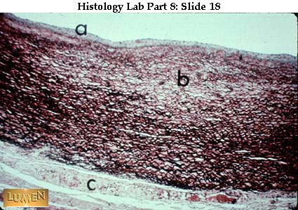
a = seminiferous tubules
b = rete testis in mediastinum
c = epididymis
d = efferent ducts Note the thick collagenous connective tissue capsule (the tunica albuginea) surrounding the testis
Higher power of rete testis, the narrow cavernous channels lying in the dense connective tissue. You will notice a couple of thin-walled, blood-filled blood vessels also coursing in the same region. Sperm produced in the seminiferous tubules leave the testis by way of the rete, which ultimately converges on about twelve efferent ducts. Detail of rete testis, showing cavernous, irregular channels lined with a low epithelium. Low power view of seminiferous tubules. These tubules are very long and tightly coiled, so each one is cut many times in any given section of the testis. Blood vessels and interstitial cells of Leydig lie in the connective tissue stroma between the tubules. |
| | | | ابراهيم الشنطي
Admin
عدد المساهمات : 70301
تاريخ التسجيل : 28/01/2013
العمر : 77
الموقع : الاردن
| | موضوع: رد: صور طبية هيستولوجى - Histology الإثنين 19 نوفمبر 2018, 12:18 am | |
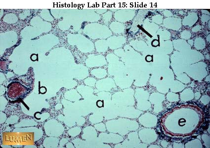
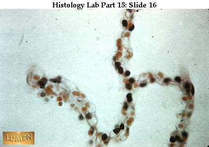
| Female Reproductive Tract - Cervix, Vagina, Umbilical Cord, Placenta, and Mammary Gland  Mucosa of the cervix with its lumen to the left. (The uterus would lie above this region and the vagina below.) Notice how the mucosal glands slant upwards. They produce a mucoid secretion. Arrows = small blood vessels. ــــــــــــــــــــــــــــــــــــــــــــــــــــــــــــــــــــــــــــــــــــــــــــــــــــــــــــــــــــــــــــــــــــــــــــــــــــــــــــ Sharp transition from simple columnar epithelium of the endocervix to non-cornified stratified squamous epithelium of the ectocervix and vagina. ـــــــــــــــــــــــــــــــــــــــــــــــــــــــــــــــــــــــــــــــــــــــــــــــــــــــــــــــــــــــــــــــــــــــــــــــــــــــــــــ Vagina with stratified squamous epithelial lining and a wide lamina propria (some people would call the deeper portion of this layer the submucosa. The two connective tissue Iayers merge because there is no muscularis mucosae to separate them.) Notice distended venules in the connective tissue and the way the smooth muscle of the muscularis externa lies in loose strands. How would you distinguish this slide from a slide of esophagus? ـــــــــــــــــــــــــــــــــــــــــــــــــــــــــــــــــــــــــــــــــــــــــــــــــــــــــــــــــــــــــــــــــــــــــــــــــــــــــــ Vaginal smear taken during the reproductive years when the epithelium is thickest. Specifically, this smear was taken on day nine of the menstrual cycle. The large squames were removed from the surface of mature stratified squamous epithelium. ــــــــــــــــــــــــــــــــــــــــــــــــــــــــــــــــــــــــــــــــــــــــــــــــــــــــــــــــــــــــــــــــــــــــــــــــــــــــــــــ This smear was taken on day 25 of the menstrual cycle and also shows the squames of mature stratified squamous epithelium. The numerous rod- like structures in the field are lactobacilli. ـــــــــــــــــــــــــــــــــــــــــــــــــــــــــــــــــــــــــــــــــــــــــــــــــــــــــــــــــــــــــــــــــــــــــــــــــــــــــــــــــــ Vaginal smear characteristically seen before puberty and after menopause, when the surface epithelium is relatively thin, either immature or atrophic. Since there are no surface squamous cells, the smear contains primarily rounded parabasal cells. Notice the presence of numerous small neutrophils with their "beady", dense, lobulated nuclei. The vagina is more prone to infections when the epithelial lining is thin. ــــــــــــــــــــــــــــــــــــــــــــــــــــــــــــــــــــــــــــــــــــــــــــــــــــــــــــــــــــــــــــــــــــــــــــــــــــــــــــــــــ Both of these smears contain endocervical epithelial cells of a simple columnar, mucus-secreting type. ــــــــــــــــــــــــــــــــــــــــــــــــــــــــــــــــــــــــــــــــــــــــــــــــــــــــــــــــــــــــــــــــــــــــــــ Again, these are endocervical cells, but this time they are a twisted strand of ciliated epithelium. ــــــــــــــــــــــــــــــــــــــــــــــــــــــــــــــــــــــــــــــــــــــــــــــــــــــــــــــــــــــــــــــــــــــــــــــــــــــــــــــــــــــــ One of the umbilical arteries with its typically thick coat of smooth muscle. The lumen is filled with blood. Surrounding the vessel is the pale gelatinous Wharton's jelly, a specialized connective tissue with very few cells and hardly any fibers, but lots of viscous ground substance. This "jelly" contributes to the "rubberiness" of the umbilical cord, making it quite turgid and therefore unlikely to tie itself into hard knots or strangle the fetus. The umbilical cord contains two arteries and one vein. ـــــــــــــــــــــــــــــــــــــــــــــــــــــــــــــــــــــــــــــــــــــــــــــــــــــــــــــــــــــــــــــــــــــــــــــــــــــــــــــ  Low power view of the "fetal" surface of the placenta. The right-hand surface is the thick, pale pink chorionic plate. Extending to the left, from the plate, are some large, pale pink, chorionic stem villi which are seen giving off branches along their edges. All the fine pieces of tissue making up the bulk of the placenta are actually the smallest branches of the chorionic villi. What looks like empty space around them is really the intervillous space normally filled with maternal blood. The placenta, therefore, is primarily composed of fetal (chorionic) tissue (chorion frondosum) lying in a bath of maternal blood. The fetal blood flows in the vessels that you see lying in the substance of the chorionic plate and the chorionic villi, right out into the finest branchings. Such a placenta is termed hemochorial because the maternal blood (hemo-) is in direct contact with the fetal chorionic tissue (-chorial). ــــــــــــــــــــــــــــــــــــــــــــــــــــــــــــــــــــــــــــــــــــــــــــــــــــــــــــــــــــــــــــــــــــــــــــــــــــ Detail of thin wall of amnion, slightly detached from the chorionic plate below. The simple cuboidal epithelium of the amnion makes a smooth surface facing the fetus, which is lying in the fluid contained in the amnionic sac. ــــــــــــــــــــــــــــــــــــــــــــــــــــــــــــــــــــــــــــــــــــــــــــــــــــــــــــــــــــــــــــــــــــــــــــــــــــــــــــــ Detail of the opposite surface of the placenta, that is, the maternal side that is eroding the endometrium of the uterus. Chorionic villi at the right are seen attaching to the thick decidual plate to the left. Large decidual cells are characteristic of this plate. They represent modified endometrial stromal cells undergoing the so-called decidual reaction during pregnancy. This plate will be sloughed off as part of the "afterbirth" at the time of parturition (expulsion of the fetus). Only the basal layer of the endometrium will remain intact after birth; it will then begin rebuilding under hormonal influences. ــــــــــــــــــــــــــــــــــــــــــــــــــــــــــــــــــــــــــــــــــــــــــــــــــــــــــــــــــــــــــــــــــــــــــــــــــــــــــــــ Low power view of the nipple of a lactating cat, showing lobules of the mammary gland below and several narrow lactiferous ducts exiting upwards toward the surface of the nipple. The mammary gland is a compound alveolar gland derived originally as multiple epithelial ingrowths from the skin. The epithelium of its ducts and secretory units is directly continuous with the epidermis of the nipple area. ـــــــــــــــــــــــــــــــــــــــــــــــــــــــــــــــــــــــــــــــــــــــــــــــــــــــــــــــــــــــــــــــــــــــــــــــــــــــــــ Mammary gland (inactive): composed mostly of pale, wide, connective tissue interlobular septa with scattered lobules containing small dark cross-cuts of many intralobular ducts. There are very few, if any, secretory alveoli in the inactive gland. Much of the interlobular tissue is adipose tissue. There is one, large, interlobular duct toward the lower right corner of the field. ـــــــــــــــــــــــــــــــــــــــــــــــــــــــــــــــــــــــــــــــــــــــــــــــــــــــــــــــــــــــــــــــــــــــــــــــــــــــــــــــــــــ Detail of one lobule of an inactive mammary gland. Note that the intralobular ducts branch frequently but have no secretory acini at their endings. ـــــــــــــــــــــــــــــــــــــــــــــــــــــــــــــــــــــــــــــــــــــــــــــــــــــــــــــــــــــــــــــــــــــــ Another view of intralobular ducts of an inactive gland. Dense connective tissue and fat cells lie in the surrounding interlobular septa. The connective tissue stroma within the lobule is more cellular than the interlobular connective tissue outside. ــــــــــــــــــــــــــــــــــــــــــــــــــــــــــــــــــــــــــــــــــــــــــــــــــــــــــــــــــــــــــــــــــــــــــــــ Mammary gland (proliferating in pregnancy) with the lobules now enlarging as secretory acini sprouts from the intralobular duct systems. The septa (pale pink) are becoming compressed. Note large interlobular ducts lying in the septa. Lobules now seem more comparable to the kind of thing seen in the salivary gland. Secretion of watery cholostrum precedes the secretion of true milk, which does not come until after the birth of the child. ــــــــــــــــــــــــــــــــــــــــــــــــــــــــــــــــــــــــــــــــــــــــــــــــــــــــــــــــــــــــــــــــــــــــــــــــــــــــــ  Diagram of inter-relationships between the anterior pituitary, ovary and uterus during the menstrual cycle. Beginning at the upper left, the ovarian follicle enlarges under the influence of high titers of FSH from the pituitary. As the follicle grows, its theca interna produces increased amounts of estrogen which causes the endometrium below to thicken. During this proliferative phase, the endometrial glands are thin and straight and the coiled arteries increase in length. At mid-cycle (about 14 days) there is a great surge of LH from the pituitary, coinciding with the time of ovulation. The follicular epithelium that remains behind undergoes a marked hyperplasia and differentiates into granulosa lutein cells, which form the bulk of the new corpus luteum. Under the influence of pituitary LH, these cells now produce progesterone which, in turn, causes the endometrium to thicken somewhat further and develop very wide, tortuous, sacculated glands, ready for implantation by an ovum. Estrogen is still being produced by the theca interna. At about 28 days, if there is no implantation, the titers of estrogen and progesterone fall off as the corpus luteum degenerates, and, at the same time, the coiled arteries of the endometrium clamp down. Thus deprived of nourishment, the endometrium begins to break up and slough off in menstruation. Only the basal layer of the endometrium will remain. Hormonal feedback now tells the pituitary to increase its secretion of FSH, thus starting the cycle all over again. In the event of pregnancy, of course, the corpus luteum is preserved, the production of estrogen and progesterone remains high, and the glandular endometrium is maintained. In time, the developing placenta itself produces an LH-like chorionic gonadotropin and, later, both estrogen and progesterone, in order to maintain the appropriate hormonal environment for the developing fetus and its needs. ـــــــــــــــــــــــــــــــــــــــــــــــــــــــــــــــــــــــــــــــــــــــــــــــــــــــــــــــــــــــــــــــــــــــــــــــــــــــــــــــــــــــ Detail of secretory acini of the proliferating mammary gland. An intrabular blood vessel is evident in the upper left quadrant of the field. ــــــــــــــــــــــــــــــــــــــــــــــــــــــــــــــــــــــــــــــــــــــــــــــــــــــــــــــــــــــــــــــــــــــــــــــــــــــــــ Lactating mammary gland with alveoli (acini) very distended with milk secretion, which stains bright pink here. Notice the branching, tubular shapes to some of the secretory units. The lobule to the right has emptied its contents. Notice how thin and compressed the interlobular connective tissue septa are now (very thin, pink strands around groups of the empty alveoli). |
| | | | ابراهيم الشنطي
Admin
عدد المساهمات : 70301
تاريخ التسجيل : 28/01/2013
العمر : 77
الموقع : الاردن
| | موضوع: رد: صور طبية هيستولوجى - Histology الإثنين 19 نوفمبر 2018, 12:18 am | |
| Female Reproducive Tract - Ovary, Oviduct, and Uterus Ovary with surface cuboidal epithelium. (Really a modified mesothelium.) ــــــــــــــــــــــــــــــــــــــــــــــــــــــــــــــــــــــــــــــــــــــــــــــــــــــــــــــــــــــــــــــــــــــــــــــــــــــــــــــــــ Cortex of ovary. A thick connective tissue capsule, the tunica albuginea underlies the surface epithelium. Somewhat deeper lie several small, primary (primordial) follicles. (All egg cells have reached the primary oocyte stage by birth and are held in this "suspended animation", in very early prophase, until such time as they may ovulate or undergo atresia.) ــــــــــــــــــــــــــــــــــــــــــــــــــــــــــــــــــــــــــــــــــــــــــــــــــــــــــــــــــــــــــــــــــــــــــــــــــــــــ Primary follicles with one single layer of flat follicle cells surrounding an oocyte. Although an oocyte is a giant compared with its neighbors, this early stage is small for an oocyte, and the cell will grow considerably in size when it begins to mature, under the influence of FSH. The nucleus looks lightly granular, and the dark nucleolus is prominent. Cytoplasm is very pale. Note the "swirly" interstitial tissue of the ovarian stroma. ــــــــــــــــــــــــــــــــــــــــــــــــــــــــــــــــــــــــــــــــــــــــــــــــــــــــــــــــــــــــــــــــــــــــــــــــــــــــــــــــــــــ Early maturation stage of follicle with beginning proliferation of follicle cells around an enlarging oocyte. The nucleolus shows clearly inside the nucleus. As the oocyte enlarges, its chromosomes prepare further for the first meiotic division, which will occur at ovulation. ــــــــــــــــــــــــــــــــــــــــــــــــــــــــــــــــــــــــــــــــــــــــــــــــــــــــــــــــــــــــــــــــــــــــــــــــ Further developed follicle
- a = with antrum beginning at arrows. The homogenous gray-blue line immediately surrounding the egg cell itself is the zona pellucida.
- b and c = primary follicles, containing oocytes which are still small.
ـــــــــــــــــــــــــــــــــــــــــــــــــــــــــــــــــــــــــــــــــــــــــــــــــــــــــــــــــــــــــــــــــــــــــــــــــــــــــــ A group of follicles in various stages of early development in the cortex of a rat ovary. Blood vessels of the ovarian medulla are seen in the center of the field. Development of follicles is regulated by FSH from the anterior pituitary. ــــــــــــــــــــــــــــــــــــــــــــــــــــــــــــــــــــــــــــــــــــــــــــــــــــــــــــــــــــــــــــــــــــــــــــــــــــ  Maturing follicle, so called because it contains a definite antrum (or fluid-filled space) and many layers of granulosa cells. The egg is still a primary oocyte and sits to one side of the follicle on a mound of cells called the egg hillock or cumulus oophorus. The cells closest to the oocyte will be expelled with it at ovulation as the corona radiata. Surrounding the granulosa cells of the follicle is the theca interna, a rather cellular and vascular connective tissue layer, which secretes estrogen. Outside of this is the theca externa a more fibrous connective tissue layer, not well defined here. Note that several follicles may start to develop in any one monthly cycle, but in the human only one will mature, unless there are to be multiple ovulations and therefore possible multiple births. All follicles that don't complete their maturation undergo atresia (i.e., degenerate). The egg dies, the granulosa layer breaks up, and the whole follicle collapses and undergoes fibrotic change. ــــــــــــــــــــــــــــــــــــــــــــــــــــــــــــــــــــــــــــــــــــــــــــــــــــــــــــــــــــــــــــــــــــــــــــــــــــــــــ Large ruptured follicle, just after ovulation.
- Arrow = stigma, the point of rupture where oocyte was expelled. The reduction division (or first meiotic division) takes place at the time of ovulation.
- a = granulosa cells that will now proliferate under the stimulus of pituitary LH and enlarge to become granulosa lutein cells, filling in the follicular cavity and becoming the major portion of the new corpus luteum.
- b = corpus albicans -- old scar of an earlier corpus luteum
ــــــــــــــــــــــــــــــــــــــــــــــــــــــــــــــــــــــــــــــــــــــــــــــــــــــــــــــــــــــــــــــــــــــــــــــــــــــــــــــــــــــــــ Detail of corpus luteum showing the rounded foldings of large, pale granulosa lutein cells in the lower half of the picture; these secrete progesterone. Pushing down between the folds is a wedge of smaller, darker theca lutein ; these secrete estrogen. ــــــــــــــــــــــــــــــــــــــــــــــــــــــــــــــــــــــــــــــــــــــــــــــــــــــــــــــــــــــــــــــــــــــــــــــــــــــ Detail of granulosa lutein cells with very large, pale steroid secreting cells. Their nuclei are round and granular looking, and each contains a dark, prominent nucleolus. Cell cytoplasm should be pale pink (overstained with hematoxylin here). With EM, the cytoplasm would show abundant smooth ER, as is typical of steroid secreting cells. ــــــــــــــــــــــــــــــــــــــــــــــــــــــــــــــــــــــــــــــــــــــــــــــــــــــــــــــــــــــــــــــــــــــــــــــــ Corpus albicans, which is a scar of dense collagenous (white fibrous) tissue; the remnant of a degenerated corpus luteum. ـــــــــــــــــــــــــــــــــــــــــــــــــــــــــــــــــــــــــــــــــــــــــــــــــــــــــــــــــــــــــــــــــــــــــــــــــــــــــــــــــــــ Detail of corpus albicans. Nuclei represent fibroblasts. ــــــــــــــــــــــــــــــــــــــــــــــــــــــــــــــــــــــــــــــــــــــــــــــــــــــــــــــــــــــــــــــــــــــــــــــــــــــــــــــــــــــ Oviduct with highly labyrinthine mucosa. Each piece of folded, branching mucosa is lined with simple columnar epithelium. The rest of the wall is rather thin and shows interlaced smooth muscle bundles. ــــــــــــــــــــــــــــــــــــــــــــــــــــــــــــــــــــــــــــــــــــــــــــــــــــــــــــــــــــــــــــــــــــــــــــــــــــــــــ A higher power of the fimbriated (finger-like) end of the oviduct. The surface epithelium is high cuboidal or low columnar and has a ciliated surface. Arrows indicate non-ciliated "peg" cells which are secretory in function and stand up higher than the other cells. (a = lamina propria core of fimbria.) ــــــــــــــــــــــــــــــــــــــــــــــــــــــــــــــــــــــــــــــــــــــــــــــــــــــــــــــــــــــــــــــــــــــــــــــــــــــــــ A panoramic view of the uterus showing the whole thickness of the wall.
- a = endometrium (a proportionally thin layer, with a dark base as seen here.) This is a mucosa, with epithelium and lamina propria and glands.
- b = wide, dark myometrium (smooth muscle in irregular, spiralling layers). This is by far the widest layer in the wall.
- c = connective tissue perimetrium (adventitia).
- Arrows point to large blood vessels
ــــــــــــــــــــــــــــــــــــــــــــــــــــــــــــــــــــــــــــــــــــــــــــــــــــــــــــــــــــــــــــــــــــــــــــــــــــــــ  Dotted line represents the base of the endometrium. Menstruation is in progress, with breakdown of surface tissue. ــــــــــــــــــــــــــــــــــــــــــــــــــــــــــــــــــــــــــــــــــــــــــــــــــــــــــــــــــــــــــــــــــــــــــــــــــــــــــــ Overview of uterine wall in the early post-menstrual stage. Only the basal layer of endometrium is present. Glands are sparse. ــــــــــــــــــــــــــــــــــــــــــــــــــــــــــــــــــــــــــــــــــــــــــــــــــــــــــــــــــــــــــــــــــــــــــــــــــــــــــــــــــ View of endometrium in an early proliferative stage (same magnification as the previous slide). The endometrium has grown thicker under the influence of estrogen. Glands are straight and thin. ــــــــــــــــــــــــــــــــــــــــــــــــــــــــــــــــــــــــــــــــــــــــــــــــــــــــــــــــــــــــــــــــــــــــــــــــــــــــــ View of a later proliferative stage. Glands are still quite straight and thin, but there is a noticeable difference between the darker, more compact basal and the paler, more edematous functional layer above. ـــــــــــــــــــــــــــــــــــــــــــــــــــــــــــــــــــــــــــــــــــــــــــــــــــــــــــــــــــــــــــــــــــــــــــــــــــــــــــــــ Endometrium in the early secretory stage. Glands are becoming tortuous and sacculated under the influence of progesterone. Their glycogen-rich mucoid secretion is stored within the glands, pending a possible implantation of an embryo. ـــــــــــــــــــــــــــــــــــــــــــــــــــــــــــــــــــــــــــــــــــــــــــــــــــــــــــــــــــــــــــــــــــــــــــــــــ View of later secretory stage. Sacculation of glands has increased, and menstruation seems to have begun near the surface. ــــــــــــــــــــــــــــــــــــــــــــــــــــــــــــــــــــــــــــــــــــــــــــــــــــــــــــــــــــــــــــــــــــــــــــــــــــــــــــــــ Endometrium in secretory (progestational) stage with sacculated glands. The thinner, non-sacculated bases of the glands will remain after menstruation, and their epithelial lining will undergo mitotic activity in order to provide the new surface epithelium for the uterine lumen. ــــــــــــــــــــــــــــــــــــــــــــــــــــــــــــــــــــــــــــــــــــــــــــــــــــــــــــــــــــــــــــــــــــــــــــــــــــــــــ Surface epithelium (simple cuboidal to columnar) of the uterine lining. Patches of cells are ciliated. |
| | | | ابراهيم الشنطي
Admin
عدد المساهمات : 70301
تاريخ التسجيل : 28/01/2013
العمر : 77
الموقع : الاردن
| | موضوع: رد: صور طبية هيستولوجى - Histology الإثنين 19 نوفمبر 2018, 12:19 am | |
| Stains, Cells, and Ultrastructure (EM)
In a low power view of intestinal wall, rows of epithelial nuclei impart a darker, bluer color to linings of surfaces and glands, as seen to the right of center. The outer, left-hand layers show the pink of muscle cytoplasm. The middle layer of dense, irregular connective tissue shows how brightly collagen fibers can be stained with eosin.
High power of smooth muscle to show that eosinophilic color is mainly due to cytoplasm. Nuclei are quite scattered and have only small, granular clumps of blue heterchromatin. Nucleoli (one or two per nucleus) are stained blue with hematoxylin.
Intestinal wall stained with Mallory's trichrome stain, which specifically colors collagen fibers blue. With this stain the connective tissue layer is clearly distinguished from muscle below and epithelium above, both of which take the pink/purple stain of cytoplasm.
Detail of a group of epithelial cells containing bright red (eosinophilic) secretory granules. Nuclei are dark with hematoxylin.
This organ, the thymus, appears very basophilic in H&E.
At high power, the reason for the basophilia is clear: the thymus is packed with lymphocytes with darkly stained nuclei. Isolated structures such as the whorl of cells in the center, are specifically acidophilic (eosinophilic).
Here are some nerve cells, seen in low power. Their nuclei are pale and vesicular, containing mainly unstained euchromatin. The nucleolus is dark, however, and the cvtoplasm is filled with clumps of darkly stained, basophilic material, implying a content of ribonucleic acid.
Cells take diverse shapes. These are epithelial cords of block-like cells. As always, nucleoli and nuclear heterochromatin stain darkly with hematoxylin.
Blood cells are suspended in fluid plasma and therefore are characteristically round in shape.
Muscle cells are arranged parallel to their direction of contraction and adopt a fusiform or spindle shape. Nuclei are sparse in relation to large amounts of cytoplasm.
In low power, individual muscle cell groups are found to be running in different directions, so that some are cut cross-wise (or transversely) and some are cut lengthwise (longitudinally). Some, of course, are running obliquely and therefore are cut tangentially in relation to their full length.
Nerve cells are typically stellate in shape, with several cytoplasmic extensions or processes. Here again, notice that the cytoplasm of these cells contains dark, basophilic material. In EM, this material will turn out to be abundant rough endoplasmic reticulum, which is associated with protein production. Before leaving this slide, note the many tiny nuclei in the field, in between the two nerve cells. Their size is about equal to the nucleolus of a nerve cell!
Another type of nerve cell, to show again its huge size in relation to the ordinary connective tissue cells around it. Once again, the nucleolus of the nerve cell (lying in the rather small, pink nucleus) is about equal in size to the nuclei of other cells. Look just below the nerve cell (at about the 5:30 position on a clock face) for a small capillary containing a single, quite pink erythrocyte. Figuring that the r.b.c. is about 7.5 microns in diameter, you can estimate the size of the neuron!
Silver staining is useful for a variety of purposes. Here it is used to blacken the reticular fiber network of reticular tissue.
Here silver has been deposited on nerve cells and their delicate processes in the brain.
In this instance, silver has been deposited on the intercellular substance between epithelial cells. You will notice that silver seems particularly useful for viewing very thin, fine structures which become visible when impregnated with grains of silver. Incidentally, this particular view is of the surface of mesothelium (simple squamous epithelium lining body cavities and mesentery).
Now a whole-mount of a small blood vessel has been stained with silver. The thin black vertical lines are reticular fibers running around the outside of the vessel like barrel hoops. The irregular horizontal lines, running parallel to the length of the vessel are the silvered outlines of endothelial cells. The intercellular cement has been stained black, making this surface view of the endothelium look like the pieces of a puzzle interlocked together. Cell nuclei are not visible.
 EM of a "typical" cell (hepatocyte), showing the organelles common to almost all cells of the body. Notice rod-like mitochondria (M), stacked rough endoplasmic reticulum, and electron-dense lysosomes. The small dots encrusting the rough ER are ribosomes; compare their size with the particles of glycogen, shown as black, irregular clusters. Notice also that the nucleus (N) contains very little heterochromatin, and seeming gaps along the nuclear envelope where the nuclear pores are found (We'll get back to other features of this cell when we study the liver.) B=bile canniculus; HS=hepatic sinusoid; SD=space of Disse.
Transmission electron micrograph of nucleus similar to the one in the previous figure. The nucleolus (3) shows an internal structure. The chromatin is predominately euchromatin with heterochromatin which is typically located close to the nuclear envelope and is discontinuous at the nuclear pores. Mitochondria (2) are seen in the surrounding cytoplasm.
Detailed EM of nucleolar structure, showing fibrillar (1), granular (2), and amorphous (3) portions.
A lymphocyte in late prophase. The nuclear envelope has begun to disappear and is evident in only a few places. (Arrow) CG = Chromatin granules. M = Mitochondria Rer = Rough endoplasmic reticulum.
A lymphocyte in metaphase with the chromosomes lined up on the equatorial plate. The plane of section does not include the spindle fibers. 1 = Endoplasmic reticulum. 2 = Mitochondria.
EM of the nuclear envelope. Dense chomatin material (heterochromatin) (1) is distributed along the nuclear envelope except in the region of the nuclear pores (2). 3=euchromatin; 4=smooth endoplasmic reticulum; 5=Golgi body.
Higher magnification micrograph with nucleus to the left and cytoplasm to the right. A pore in the nuclear envelope is marked by the arrow. Notice the absence of heterochromatin at the site of the pore. N = Nucleus.
EM of an oocyte with its nucleus (5) at the bottom of the micrograph. The nuclear envelope is sectioned tangentially so that nuclear pores are clearly visible (arrows). 1 = Crystalline bodies or plaques (typical of oocyte cytoplasm); 2 = Mitochondria; 3 = Multi vesicular body; 4 = Cortical granules (typical of oocyte); 5 = Nucleus.
EM showing the two dense and one pale (or lucent) layers of the ordinary cell (or plasma) membrane.
A similar membrane coated with a fuzzy-looking external glycocalvx (arrow). GA=Golgi apparatus.
Electron micrograph of the basal lamina. The portion of the basal lamina referred to as the lamina densa (1) is a thin gray line lying just outside the cell membrane. Reticular fibers (2) are associated with the lamina densa. Notice here that the basal lamina surrounds an epithelial cell. Two odd points to remember: (1) lymphatic capillaries have no basal lamina surrounding their endothelium, and (2) fat cells do have a basal lamina, which is surprising because these are connective tissue cells and shouldn't seem to need a protective layer between themselves and the surrounding connective tissue ground substance. The true origin of fat cells is open to question.
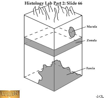 Diagram of a block-like cell showing the extent of various kinds of cell junctions. A macula is a simple "spot weld". A zonula forms a complete belt of adhesion around the cell. A fascia is a broad, irregular area of adhesion. Notice that the apical surface of the cell has several small cytoplasmic protrusions. They are like microvilli stucturally but are not numerous enough to form a striated or brush border. Such small protrusions are common on cells.
Scanning EM view looking down on the apical surface of a whole sheet of epithelial cells. The long, wavy projections are cilia: the close-cropped ones are microvilli of a brush border.
High power EM of microvilli of a brush border. Notice that they are simple extensions of the apical cytoplasm, with unit membrane continuing over their surface. Very fine actin filaments extend into the microvilli and are rooted in the main mass of cytoplasm below. Angling down the bottom half of the picture is the line of contact between two adjacent cells, each with its own unit membrane. At three points along the way there are specialized junctions: (1) zonula occludens or tight junctions, (2) zonula adherens or intermediate junction, and (3) desmosome or macula adherens. Cytoplasmic filaments (arrow) are attached to the desmosome, contributing to its density.
 Lower magnification EM of junctional complexes between epithelial cells. The tonofilaments heading into the desmosomes (5) are particularly prominent. The continuous bands of zonula occludens (3) and zonula adherens (4) are seen near the top. Note the width of the intercellular space along its normal length and at the points of various kinds of contacts. 1=microvilli; 2=terminal web.
EM detail of several desmosomes, showing the attachment of many tonofilaments. Arrows point to the density which typically appears in the intercellular space. The cell membranes of the two neighboring cells are interlocked in a very complex interdigitation here. You can follow the undulating course of the intercellular space across the picture.
EM detail of junctional complexes. In the area of the tight junction (1) (zonula occludens) the two unit membranes approach each other and appear to merge revealing only three dense lines (instead of four). In the area of the intermediate junction (2) (zonula adherens) the intercellular space narrows to about 20 nm, but there is still a space.
EM of cilia cut longitudinally. (A few microvilli are on the neighboring cell to the left, for a size comparison.) Notice that each cilium is rooted in a barrel-like basal body. The dense lines extending from the basal bodies and up into the cilia are microtubules. The unit membrane of the cell continues up over each cilium.
Cross-cuts of cilia showing the typical 9X2 +2 arrangement of microtubules within the cytoplasm ( ring of 9 doublets plus 2 single microtubules in the center). The cell membrane envelopes each cilium.
Tangential section of cilia showing the structural transitions that occur between the shaft of the cilia (upper right) and the basal bodies (lower left) which give rise to the cilia.
EM of hepatocyte illustrating size relationships between glycogen particles (1 and 2) and ribosomes of the RER (3).
Typical arrangement of cisterns of rough ER in a secretory epithelial cell. A few mitochondria (1) are at the lower right. The presence of ribosomes on the RER, with all their ribonucleic acid content, render them basophilic to stains. 2=secretory granule.
Serous secretory acini showing cytoplasmic basophilia toward their bases where a lot of rough ER lies. The presence of rough ER in such abundance signifies production of protein (in this case, some digestive enzyme). The secretory granules are pale here.
Details of a Golgi apparatus (body) showing the forming face (1); maturing face (2); saccules (3) and secretory vesicles (4) budding from the saccules. The Golgi complex typically lies adjacent to the nucleus. 5= centriole.
High magnification of a network of smooth endoplasmic reticulum. Unlike rough endoplasmic reticulum, which usually occurs in flat sheets, this organelle comprises interconnected tubules (1). 2 = Mitochondrion; 3 = Free ribosomes, seen either singly or as Polyribosomes (polysomes).
EM of microtubules, seen as fine parallel lines when cut longitudinally (lower panel) or circles when cut transversely (upper panel). Images are from dendrites and axons of neurons.
EM of plasma membrane infoldings (PF) and mitochondria (M) that are aligned parallel to the membranes. Note the basal lamina (BL) at the base of the cells.
Part of a lymphocyte showing a centriole (C) cut transversely. Note the triplet arrangement of microtubules cut in cross-section. GA = Golgi apparatus (body); PR = Polyribosomes (polysomes); NS = Perinuclear space (of the nuclear envelope).
Part of a lymphocyte showing continuity of the rough endoplasmic reticulum (rer) with the nuclear envelope (at arrow).
Cytoplasmic organelles of a renal collecting duct cell. TL = Tubular lumen; MV = Microvilli on cell surface; M = Mitochondrion; PR = Polyribosomes (polysomes); GA = Golgi apparatus (body); IS = Intercellular space; notice how corrugated the interdigitations of the cell membranes are between the two cell. (lower right).
Details of mitochondria. 1 = External envelope; 2 = Cristae; 3 = Matrix (the more electron-dense material); 4 = Granules within the matrix.
Large lipid droplets (LD) are seen within a cell in the deeper parts of the adrenal cortex. The lipid matrix has been removed during tissue preparation. M = Mitochondria (with tubular cristae are typical of steroid producing cells); SER = Smooth endoplasmic reticulum.
Detail of secondary lysosome with engulfed material within it. 1 = Limiting membrane; 2 = Matrix; 3 = partly digested material.
Different stages of the pinocytosis, an endocytic process, in an endothelial cell. The vessel lumen is to the right; the underlying connective tissue is to the left. Notice the thin gray (electron-dense) line of the basal lamina immediately along the left border of the cell. 1 = Vesicle open to the outside of cell, facing the extracellular matrix; 2 = Vesicle partially enclosed by cell membrane; 3 = Vesicle limited by membrane and wholly within cytoplasm of cell. The elongate nucleus lies in the center of the cell. |
| | | | ابراهيم الشنطي
Admin
عدد المساهمات : 70301
تاريخ التسجيل : 28/01/2013
العمر : 77
الموقع : الاردن
| | موضوع: رد: صور طبية هيستولوجى - Histology الإثنين 19 نوفمبر 2018, 12:20 am | |
| Connective Tissue Proper
Mesenchyme -- embryonic c.t. with multipotential cells. The stellate cells are beginning to form fibers. Sometimes cells are more spindle shaped. Ground substance material is watery and invisible.
Reticular tissue (silvered, black). A network of very fine reticular fibers can be seen here, forming the stroma (framework) of a lymph node. These fibers are produced by reticular cells. The pale cells seen in the meshes of the reticular fibers are lymphocytes.
Stellate reticular cells - forming a meshwork of their own cytoplasmic processes. These are in addition to the reticular fiber network which these cells produce -- and which we would see if this tissue were silvered. Notice particularly clear cells in upper left quadrant of field. This slide is from lymph node.
Detail of lymph node, showing stellate reticular cell in middle of field.
Loose (areolar) connective tissue - (in blue) - surrounding the epithelium of tubules. In areas like this, the finest collagen fibers lying closest to the tubules would be reticular fibers; the only way to distinguish them here from heavier collagen fibers would be to silver them. (The blue here simply stains collagen in general.) REMEMBER: in an area like this, reticular fibers (like all other fibers) are produced by fibroblasts. Only in the primitive reticular tissue of bone marrow, lymph node, and spleen are reticular fibers produced by reticular cells.
Loose irregular connective tissue (also called areolar tissue) as seen underlying and supporting epithelium in an ordinary section. It is rather cellular and supports many small blood vessels which travel through it.
Areolar c.t. immediately underlying simple columnar epithelium. This is a very cellular variety of areolar c.t., with a high population of lymphocytes.
A stretched preparation of areolar connective tissue. The pink fibers of different thicknesses are collagenous (or white) fibers. The dark, thin, more tortuous fibers are elastic (or yellow) fibers. Most of the nuclei belong to fibroblasts.
Dense irregular c. t., with fibers running in all directions. The fibers are mainly collagenous, but keep in mind that some would be elastic and can be seen only if specifically stained. This kind of c.t. is found where firmer packing and binding is needed. The two arrows at top of picture are pointing to elongate, dark, fibroblast nuclei.
Dense irregular c.t. (blue) packing around a nerve bundle. The coat immediately surrounding the whole nerve bundle is particularly dense and consists mainly of collagen fibers. In between the individual pale, round nerve fibers is a very fine areolar c.t. packing, with mainly reticular fibers.
Fat cells -- note nucleus and rim of cytoplasm pushed to one side by the accumulation of fat. The lipid itself has been dissolved out in fixation. In the center of the picture, in the space bounded by the four large fat cells, there is a small, round cross-cut of a capillary with a dark, shrunken red blood cell inside.
Fat cells developing in areolar connective tissue.
Adipose tissue aggregate of fat cells.
Adipose tissue as seen in a regular histological section. The pale pink tissue mixed in with it is skeletal muscle. The dark purple = serous glands. There is a small muscular artery in the middle, with a branch going off it to the left.
Tendon (dense, regular c.t.), cut longitudinally. The thick collagen fibers (pink) are lined up parallel to each other, in response to the stress placed on them by muscle and joint action. Fibroblasts are squeezed between the fibers and therefore also line up in parallel rows. We often refer to this as a "railroad train" appearance.
Tendon, cut in cross-section. The pale pink background represents the cut ends of bundles of thick collagen fibers, very closely packed together. The wispy lines you see throughout are the "cracks" between fiber bundles. In the cracks lie fibroblasts which often look triangular or stellate because of being squeezed between the fibers. |
| | | | ابراهيم الشنطي
Admin
عدد المساهمات : 70301
تاريخ التسجيل : 28/01/2013
العمر : 77
الموقع : الاردن
| | موضوع: رد: صور طبية هيستولوجى - Histology الإثنين 19 نوفمبر 2018, 12:20 am | |
| [size=32]Connective Tissue Cells[/size]
Areolar c.t. -- the thin cell running diagonally toward the lower right from the center is a fibroblast
Another fibroblast -- in the curve of the pink collagen fiber. The long, narrow nucleus is characteristic.
Several fibroblasts, lying among collagen fibers. Hardly any cytoplasm is visible.
EM of cytoplasm of fibroblast that is actively producing collagen precursors. Since collagen is a protein, we are not surprised to see a prominent rough endoplasmic reticulum. RER cisterns are packed with granular synthetic product (1). Two mitochondria (2) are visible. On the upper left-hand surface of the fibroblast, notice that secreted tropocollagen is beginning to condense into fibrillar form.
Two large macrophages (one on either side of the picture) -- with engulfed particles of blue dye in their cytoplasm. Their nuclei are pink. Compare the irregular sizes of the blue phagocytized particles here with the more even-sized granules of the mast cells in the next two slides. Notice also that the particles in the macrophage are scattered randomly.
Mast cells in areolar c.t. -- their cytoplasm full of purple secretory granules , which often seem to be spilling out. The granules contain precursors of histamine and heparin. The nuclei are hidden by the granules.
Mast cells -- deep purple metachromatic stain for granules. Again, granules are spilling out as a result of the preservation techniques. Notice how round and seed-like the granules are and how tightly they are packed in the cell. The cell nuclei are light blue.
Three large, dark mast cells in a stretched preparation of areolar connective tissue. In H & E the secretory granules stain a deep red color. Most of the other nuclei in the field belong to fibroblasts.
EM of a rat mast cell, showing typically large, homogeneously dense granules in the cytoplasm. On the left side of the micrograph, notice the presence of collagen fibrils in the extracellular space. Their presence is diagnostic for connective tissue.
EM of a human mast cell, showing a different structure for the secretory granules. Instead of being homogenous, the granules contain lamellae, whorls, and so-called paracrystalline structures. They are often described as "hair curlers" or "hair rollers"! In this picture they don't seem very densely packed, but their function seems to be very similar to that of mast cells of other species.
Plasma cell -- with somewhat basophilic cytoplasm and an eccentric nucleus with dark blocks of chromatin in it. Note the pale cytoplasmic area to the left of the nucleus; this is the negative Golgi body. Note also the pink collagen fibers scattered irregularly throughout the pale ground substance of the whole field, which is typical of areolar connective tissue.
Another plasma cell with eccentric nucleus and smooth, basophilic cytoplasm. The large, elongate, pale nucleus to the right of center belongs to a fibroblast; its cytoplasm is not visible.
Plasma cell in EM -- showing nucleolus and "cart-wheel" chromatin configuration in the nucleus. The cytoplasm is packed with rough endoplasmic reticulum, indicating protein formation. Plasma cells produce antibodies (immunoglobulins).
Eosinophils (bright pink granules) -- in areolar connective tissue. Note the bilobed nucleus in the center cell. Pale oval nuclei in the upper left hand corner probably belong to fibroblasts. Small, dark, round nuclei, such as in the lower right quadrant, probably belong to lymphocytes. Macrophages are hard to identify unless their cytoplasm is filled with phagocytized particles.
Wandering tissue eosinophils (bright pink cytoplasmic granules) and neutrophils (with lobed nuclei) -- in areolar connective tissue.
Miscellaneous cells in areolar connective tissue. The central cluster with beady, dark nuclei are wandering neutrophils. Any small, round, dark nucleus with no visible surrounding cytoplasm is a lymphocyte. The large, pale, oval nuclei scattered around the field belong to fibroblasts. In the lower right corner are two fairly oval plasma cells with definite cytoplasm and dark, round, eccentric nuclei. Macrophages are probably in the area but are hard to identify without ingested particles to mark them; the most likely candidate here is a fairly large cell just left of the central cluster with definite cytoplasm and a small oval nucleus. The rounded space in upper right corner of the field is a blood vessel with endothelial cells lining it; inside the lumen is a neutrophil, showing a dark bilobed nucleus. |
| | | | ابراهيم الشنطي
Admin
عدد المساهمات : 70301
تاريخ التسجيل : 28/01/2013
العمر : 77
الموقع : الاردن
| | موضوع: رد: صور طبية هيستولوجى - Histology الإثنين 19 نوفمبر 2018, 12:21 am | |
| Specialized Connective Tissue: Cartilage and Bone
Hyaline cartilage (lavender matrix), with perichondrium (pink) outside it. The latter is a dense regular collagenous c.t.. There are collagenous and elastic fibers lying in the cartilage matrix but they are invisible because their refractive index is the same as that of the matrix. Cartilage cells = chondrocytes, and they are lying in the lacunae.
Two chondrocytes completely filling their lacunae. If the cells were to drop out, you would see spaces in the matrix. The matrix appears very smooth, clear, and glassy (or "hyaline").
Electron micrograph of a chondrocyte in its lacuna and almost entirely filling the lacunar space. Notice that the cell has many fine cytoplasmic projections when viewed by electron microscopy. There are surrounded by heavily condensed ground substance which appears less dense on the other side of the cell.
Appositional growth of cartilage by conversion of long, thin perichondrial cells (at the right) into the round, large chondrocytes. Notice how they change shape as they lay matrix down around themselves. The cells of the outer perichondrium are fibroblasts; the inner perichondrial cells include some primitive connective tissue cells which differentiate into chondroblasts and then into chondrocytes as they lay down matrix and become embedded in it.
Hyaline cartilage with quite basophilic matrix immediately surrounding the lacunae. Cells are often grouped in "nests" (or isogenous groups) as a result of earlier mitoses and nowhere for cells to move apart. (This is called interstitial growth). (The "ripple lines" in the matrix here are due to uneven cutting of the section.)
Elastic cartilage, with chondrocytes and matrix as before, but elastic fibers predominate and take a specific stain. They always look very distinct and dark and show many branchings.
More elastic cartilage. The matrix immediately surrounding each cell is typically not traversed by fibers.
Fibrocartilage, with wispy, broad collagenic fibers predominating in the matrix. They look "cotton-y", unlike the sharply defined elastic fibers seen before. Notice that the cells are lying in lacunae.
Fibrocartilage, at the point of junction between hyaline cartilage (lavender) above and dense collagenous tissue (pink) below. The combination of chondrocytes, matrix, and visible wispy collagenic strands or fibers identifies this as fibrocartilage.
Section of compact ground bone - dry and unstained - showing cross-cuts of Haversian systems. In the center of each system is an Haversian canal which carries blood vessels. With so many such systems per unit volume of bone, we can say that bone is a well vascularized tissue. (By contrast, cartilage is avascular.)
Higher power of ground compact bone. You can see on the left that a central vascular channel (Haversian canal) is surrounded by concentric lamellae (layers) of bone. These lamellae are made up of collagenous fibers and inorganic salt matrix. The lamellae in the center of the picture are interstitial lamellae, left over from earlier Haversian systems that have been partially resorbed as new systems were laid down during the constant remodelling of the bone as it formed. Black spaces air-filled lacunae in which osteocytes once lived.
Detail of Haversian system, showing the tiny, spidery canaliculi extending from one lacuna to the next. In life these canaliculi held the processes of osteocytes thus permitting diffusion of nutrients from the central blood vessels to the outer lamellae of the Haversian system.
Detail of lacuna, showing radiating canaliculi. Tissue fluid from the capillaries and connective tissue of the Haversian canal can seep through these spaces and channels, bringing nutrients to the stellate osteocytes residing there.
EM of osteocyte in lacuna. The cytoplasm of the cell contains rough endoplasmic reticulum for the production of protein collagen, some of which can be seen lying immediately around the cell. The collagen becomes masked by black apatite (CaPO4) crystals as the matrix becomes mineralized.
High power EM of contact between two neighboring osteocytes whose processes have met in a canaliculus. Close examination of the contact shows fused outer leaflets of cell membrane (note three dark lines), indicating that this is a tight junction. Osteocytes are also known to make contact by means of gap junctions.
Low power view of a cross-cut shaft of decalcified long bone. The bone itself is pink and lies in the center of the field. The pinkness is due to the staining of collagen fibers in the lamellae. To the left is bone marrow; to the right is attaching skeletal muscle.
Early compact bone, decalcified so it can be stained. This has been cut so that the Haversian systems are cut in cross section. Vascular channels cut longitudinally are parts of Volkmann's canals.
Vascular elements from bone marrow (on the left) are continuous with vascular spaces within the bone. The endosteum lining the marrow cavity is therefore continuous with the endosteal linning of Haversian canals.
Detail of bone-forming osteoblasts lined up along the inner (endosteal) edge of bone next to the marrow cavity. In young bones growth continues in width, constantly laying down bone and resorbing it and laying down more. Real width, of course, increases by the laying down of periosteal bone on the outside of the bone, but activity continues on the endosteal surface also. Notice osteocytes inside the bony substance, lying in lacunae.
Detail of osteocytes in lacunae. The collagenous fibers of the decalcified matrix are quite acidophilic, as always. Osteocytes like these are present in both compact and spongy bone; their arrangement, however, is in concentric lamellae in compact bone and in randomly arranged lamellae in spongy bone. Remember, too, that osteocytes have processes which extend out into canaliculi in both kinds of bone. |
| | | | ابراهيم الشنطي
Admin
عدد المساهمات : 70301
تاريخ التسجيل : 28/01/2013
العمر : 77
الموقع : الاردن
| | موضوع: رد: صور طبية هيستولوجى - Histology الإثنين 19 نوفمبر 2018, 12:25 am | |
| [size=32]Endochondral Ossification[/size]
All of the long bones and many others of the body, are preformed embryologically in hyaline cartilage and then replaced by bone by endochondral ossification. Such a change has begun in the middle of the shaft of this bone, thanks to the invasion of blood vessels and their accompanying primitive connective tissue. Pale pink cartilage is seen in the head of the bone. A dark pink periosteal bone collar has already formed around the middle of the shaft, and ossification is proceeding toward both ends of the cartilage model. The dark pink strands lying outside the whole bone are dense collagenous tissue of periosteum (around the bony part) and perichondrium (around the cartilaginous part). H & E stain.
 Endochondral ossification in greater detail. The cartilage cells (chondrocytes) near the region of active ossification have enlarged (hypertrophied) and lined up more or less in columns. The purplish material in the center of the shaft is primitive bone marrow, with reticular cells and developing blood cells. The vascular elements of the marrow tissue actively invade the cartilage above, leaving spicules of calcified cartilage, upon which bony matrix will be deposited. The dark pink spicules here are made of bone; the paler pink, small spicules at the leading edge of the cartilage are made of calcified cartilage.
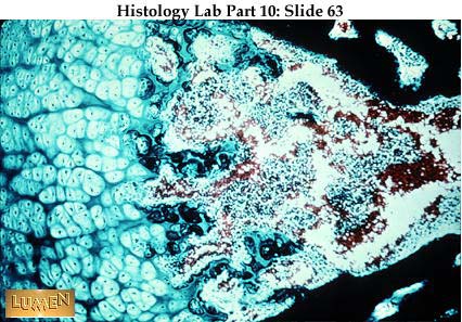 Endochondral ossification in Mallory stain. Cartilage is light blue and bone is dark blue. A thin layer of bone has already been laid down on the surface of the cartilage spicules along the leading edge of cartilage. Blood cells in the marrow cavity are red. The very dark blue at the lower left and right is spongy bone of the periosteal bone collar of the shaft. This will later be remodeled into Haversian systems of compact bone.
Head of fetal bone still made of hyaline cartilage. Near the point where ossification is going on (upper right corner) the cartilage cells become larger and the cartilage matrix becomes calcified (purple instead of pale pink here, as stained in H & E). A small amount of dark pink bone has been laid down on the surface of the calcified cartilage. Later on, a secondary center of ossification will form in the head of the bone, and the cartilage that remains between the two centers of ossification will be the epiphyseal plate for growth of the bone in length.
Region of ossification at higher magnification -- same stain as previous slide. Chondrocytes are hypertrophied, degenerating, and lined up in columns at the right. As the marrow tissue invades the cell columns, spicules of cartilage will be left. The cartilage matrix is calcified (purple), and one small area of bone deposition, has begun on it (the red color at the upper right). The small cells caught in the red matrix are osteocytes.
Another detail of ossification. Calcified cartilage spicules are purple-blue; bone deposits are purple-red. Gradually the cartilaginous portions will be resorbed as the bone is constantly reshaped, until finally there will be no trace of cartilage left. The main purpose of the cartilage in the first place was to provide a framework upon which bone deposition could begin.
Spicules showing early endochondral ossification. In H & E stain, the centers of the spicules show the purple of calcified cartilage; the edges are pink because of the bony matrix laid down upon the cartilage.
Spicules of spongy bone (bright red) surrounded by a whole line-up of osteoblasts. The osteoblasts that have previously been trapped in their own salt deposits now lie in lacunae within the spicule and are called osteocytes. The cells of the primitive bone marrow lie between bone spicules.
Detail of bony spicule with typically acidophilic (pink in H & E) matrix. Osteoblasts are lined up along its borders, depositing another layer of matrix. Osteocytes lie within lacunae in the spicule.
EM of active osteoblast laying down the fibers and salts of bone. The cytoplasm of the cell is to the left and contains lots of rough endoplasmic reticulum and many mitochondria. In the lower right corner is mineralized bony matrix containing the typical black CaPO4 (apatite) crystals. Between this matrix and the osteoblast lies a pale area of newly secreted pre-bone (or osteoid) which contains collagen fibrils (note their cross-striations) lying in an as yet unmineralized ground substance.
Spicules of spongy bone stained with Mallory stain. Bone stains blue. Note line of osteoblasts along left hand edge.
Enlarged area of spongy bone, so called because there are large, irregular spaces of bone marrow intermixed with the bony substance.
The large central space is a resorption area where young compact bone is being actively remodeled. This is an area where osteoclasts are resorbing the bony substance; notice to large multinucleated osteoclasts toward the left of the cavity next to the intact Haversian system that lies in the upper left corner of the field. Later, osteoblasts will differentiate from the primitive reticular tissue in the resorption cavity and will begin to lay down new bony lamellae around the edge of the cavity. As successive lamellae are laid down, the cavity will gradually grow smaller, until eventually a new Haversian system with a narrow central canal will be formed.
Detail of an osteoclast, a giant, multinucleated cell associated with bone resorption. The shallow bay in which it lies is a Howship's lacuna. The osteoclast is now considered to develop from a separate stem cell in the bone marrow.
EM of an osteoclast, with its ruffled border next to the area where bony matrix is being resorbed. The net effect of a ruffled border is to increase the cell surface area for contact with the collagen fibrils and apatite crystals being resorbed.
Haversian systems of decalcified compact bone, mostly cut in cross section here. The one channel cut longitudinally is a Volkmann's canal; these channels run perpendicular to both the long axis of the bone and the central canals of the Haversian systems.
Decalcified bone with Haversian system cut longitudinally. Notice the central blood vessel and the many concentric bony lamellae around it. As always, osteocytes are trapped in their lacunae.
End of a young long bone, with the pale epiphyseal plate lying between the primary ossification center of the shaft and the secondary ossification center of the head. The plate and the pale area continuous with it, up over the head, are composed of hyaline cartilage. Active ossification is going on along the lower edge of the epiphyseal plate, allowing growth in length of the bone. There is also active ossification along the lower edge of the cartilage that surrounds the head, thus allowing for growth in size of the head of the bone. Bony spicules are seen throughout the centers of ossification, making areas of spongy bone with red marrow between the spicules.
 Diagram of a cross-cut chunk of wall of the shaft of a long bone. Most of the substance is compact bone, with Haversian systems cut cross-wise on the uppermost surface and longitudinally on the right-side surface. Volkmann's canals carry blood vessels from the inner and outer bone surfaces to the vessels of the Haversian canals. The lamellae of the Haversian systems are pulled out here so that you can see the lamellar rings. External (or periosteal) circumferential lamellae are seen surrounding the whole bone. Internal (or endosteal) lamellae line the inner surface next to the marrow cavity (to the left). Notice that the inner, endosteal wall bears many spicules of spongy (cancellous, trabecular) bone. The dense collagenous connective tissue coat (the periosteum) looks dark here and surrounds the whole shaft. |
| | | | ابراهيم الشنطي
Admin
عدد المساهمات : 70301
تاريخ التسجيل : 28/01/2013
العمر : 77
الموقع : الاردن
| | موضوع: رد: صور طبية هيستولوجى - Histology الإثنين 19 نوفمبر 2018, 12:26 am | |
| Bone Marrow and Hemopoiesis
Bone marrow seen with low power (to the left). This marrow cavity lies within a shaft of compact bone (middle, pink band). Attaching muscle is to the right.
Bone marrow surrounding a pink, Y-shaped piece of spongy bone. Notice the small osteocytes scattered within the bony matrix. In the marrow there are clumps of small blood-forming cells scattered among the large round fat cells. (The lipid content of the fat cells has been dissolved out in fixation of the tissue). The elements formed most abundantly in marrow are r.b.c.'s, granular leukocytes, and platelets. Lymphocytes and monocytes are also formed here, but go elsewhere to proliferate. Another name for blood-forming tissue such as this is hemopoietic or myeloid tissue.
Bone marrow showing the typical cellular masses of developing blood cells lying between the round, empty fat cells. There are two large megakaryocytes in the field, one just about in the center and the other to the extreme right. Notice orange-colored rbc's in thin-walled sinusoids.
Bone marrow - higher power - identifiable by the fat cells, clusters of developing blood cells, and the large megakaryocytes. Also, in the middle of the field is a cross section of a sinusoid filled with rbc's.
Sinusoid of bone marrow seen here in longitudinal section. There is a good nucleus of a lining endothelial cell near the lower center of the field. Junctions between lining cells are loose so that newly formed blood cells can enter the vessels.
Megakaryocyte as seen in an H & E stained section. Note its multilobed nucleus and its comparatively giant cell size. (Remember that the other giant cell of bone, the osteoclast, has multiple separate nuclei. The osteoclast lies next to bone, while the megakaryocyte lies out in the middle of the marrow.)
Another megakaryocyte, this time as seen in a marrow smear with the May-Grunwald-Giemsa blood stain. In a smear the whole cell is here, though somewhat flattened. The lobed nucleus seems drawn together into a compact mass. Fragments of cytoplasm will form platelets.
In a section like this, stained with H & E, the developing blood cells are hard to identify. However, about in the middle of the field one can recognize a nearly mature eosinophil with bright red granules.
EM of eosinophil. Notice the crystalloid bar in the granules.
Identification of cells is somewhat easier in marrow stained with phloxine - methylene blue - azur II. Here we see a megakaryocyte near the rim of the fat cell to the left. Immediately below is a brightly stained eosinophil. The pale oval nucleus just to the right of the eosinophil belongs either to a reticular cell or a hemocytoblast (stem cell); both are primitive cells and similar in appearance in a section like this.
A reminder that bone marrow is one place where you find a stroma of reticular tissue. Here the tissue has been silvered so that you can see the network of fine reticular fibers that support all the blood forming cells. Large spaces represent fat cells.
Diagrammatic summary of the events that take place during maturation of red blood cells (erythropoiesis). Staining is with special blood stains (Giemsa, etc.). Primitive status is on the left; mature status is on the right.
Top line: there's a decrease in cell size (from left to right) and a decrease in basophilia (blueness) of cytoplasm. At the same time, hemoglobin increases, making the cytoplasm more and more acidophilic (pink). Basophilia is due to presence of abundant polyribosomes.
Second line: there's a decrease in nuclear size and ultimately extrusion and loss of the nucleus.
Third line: there's increased condensation of nuclear chromatin and eventual pyknosis of the nucleus (very dark, compact, dying). Also the nucleoli, evident at first, are soon lost
Maturational stages in development of granular leukocytes:
- Extreme left - myeloblast (the most primitive stage)
- Next - promyelocyte (these first two stages are undifferentiated precursors of all three granulocyte types)
- Next four: myelocyte, metamyelocyte, band cell, and mature cell
The top row represents the eosinophilic cell line, the middle row represents the neutrophilic line, and the bottom row represents the basophilic line. Note the decrease in cell size, the decrease in cytoplasmic basophilia (meaning decrease in polyribosomes), the increase in cytoplasmic granules (these first become specific and distinguishable as eosinophilic or basophilic at the myelocyte stage), and an increase in lobulation of the nucleus. The next few slides are of smears of bone marrow stained with modified Giemsa stains, so we can rely on the color of cells in identifying their developmental status
The cells shown here are all stages in the development of erythrocytes. Generally in the red blood cell line: (1) the cells become progressively smaller, (2) the cytoplasm changes from blue to pink, and (3) the nucleus becomes smaller and more condensed and ultimately is lost altogether. Cells shown here include (in developmental order):
- Top cell - proerythroblast
- Lower row
- left = basophilic normoblast or erythroblast. It is still blue, but is smaller; the nucleus is more condensed
- middle = polychromatophilic normoblast or erythroblast. Cytoplasm is grayer or muddier; nucleus is even more condensed.
- right = orthochromatic (or eosinophilic) normoblast. Cytoplasm is pinker and cell is smaller; nucleus is pyknotic
Reticulocytes with polyribosomal remnants (RNA) staining dark in their cytoplasm. They are slightly larger than the completely mature erythrocytes and are often found in the peripheral bloodstream at times when blood cells are being formed unusually rapidly (as during or after certain blood diseases). Remember not to confuse reticulocytes of the blood with reticular cells of connective tissue!
The large cell with blue cytoplasm is a "blast" cell although simple observation cannot tell us whether it's a myeloblast or an erythroblast. To the left is a group of neutrophilic band cells; the Iower two are probably more advanced, judging by their more segmented appearance. At bottom center is an orthochromatic normoblast with pyknotic nucleus. In the upper right corner (and probably lower left corner) is a lymphocyte.
 There is a large "blast" cell in the upper left group and a large promyelocyte at upper center. The latter is recognizable by the non-specific azurophilic granules in its cytoplasm, foretelling that it is heading toward one of the granulocyte lines. A basophilic normoblast with blue cytoplasm is in lower center. To the right of it are two early orthochromatic normoblasts. At bottom center is a late polychromatophilic normoblast with muddy cytoplasm. A slightly younger polychromatophilic cell is in the extreme lower left corner, with a slightly larger and less condensed nucleus. A neutrophilic metamyelocyte with indented nucleus also lies near the lower edge of the field. NOTE: Remember in our terminology that:
- erythroblast and normoblast are used interchangeably - as in basophilic erythroblast or basophilic normoblast.
- orthochromatic, orthochromic, acidophilic, eosinophilic are all used interchangeably as applied to the red cell line. Of course, eosinophilic as applied to the granulocytes refers strictly to those cells that are becoming eosinophils
|
| | | | ابراهيم الشنطي
Admin
عدد المساهمات : 70301
تاريخ التسجيل : 28/01/2013
العمر : 77
الموقع : الاردن
| | موضوع: رد: صور طبية هيستولوجى - Histology الإثنين 19 نوفمبر 2018, 12:26 am | |
| Lymphoid Tissues and Organs
This slide shows an isolated, single primary lymph nodule in the wall of the stomach stained dark purple because of the small lymphocyte accumulation. Such nodules are often found in the wall of tracts that exit to the outside and represent a first line of defense against foreign substances that might enter the body via these routes. Nodules like this in the GI tract are thought to be one source of B-type lymphocytes producing IgA antibodies.
Sometimes, as in this tonsil lying at the entrance to the GI tract, nodules are scattered within an otherwise diffuse mass of lymphoid tissue. In this particular field the nodules are secondary nodules, each containing a pale germinal center and a dark peripheral ring of cells.
Detail of secondary nodule. The germinal center is pale because of the presence of lymphoblasts and large lymphocytes with larger, paler nuclei. The small lymphocytes typical of circulating blood, with their smaller, darker nuclei, are pushed to the rim of the nodule, as are the plasma cells differentiating in the area.
Concentration of small lymphocytes, as found in densely packed lymphoid areas. You are essentially seeing just their nuclei.
Small lymphocytes intermixed with larger cells, many of them unidentifiable in a section such as this.
Survey EM of lymph node from a rat. 1=small lymphocytes; 2= medium-sized lymphocytes; 3= macrophage with particles in secondary lysosomes; 4= blood capillary with endothelial cell lining.
Various cell types found within red pulp in the rat spleen.
- 1= lymphocyte with relatively inactive looking cytoplasm.
- 2= plasma cell with its extensive system of RER.
- 3= monocyte containing Golgi body (4), centrioles (5) and small azurophilic granules (6)
- 7= extravasated red blood cell
- 8= reticular cell showing a bundle of filaments in a cell process (arrow
 Low power view of lymph node. The darker cortex at the periphery of the node has nodules, some of which show pale, mitotically active germinal centers. In the middle of the lymph node is the medulla with its dark cords of dense lymphocyte population. The medullary cords are surrounded by paler lymph channels (the medullary sinuses) which have relatively fewer lymphocytes. Lymphatic afferent vessels enter the node at the periphery, through the capsule (blue c.t. here), pouring lymph into the sinus system of the node. The lymph "percolates" through the cortical and medullary sinuses and leaves via the efferent lymphatics near the hilus of the node. While looking at this picture, be reminded that the nodules of the outer cortex and the cords of the medulla are included among the "homing areas" for B-cells, plasma cells, and helper T-cells. The thymic dependent area, where the majority of T-cells are found, is the deeper part of the cortex, seen here as the dense, diffuse lymphoid area lying below the cortical nodules and above the medullary cords. It is within this inner cortex that the postcapillarv venules also lie; they have an unusually high endothelium, and it is through their walls that lymphocytes of the bloodstream enter the substance of the node.
 Detail of outer surface of lymph node, with pale c.t. capsule on the outside, sending an extension (trabecula) down into the substance of the node as a frame-work. Pale sinus channels can be seen lying immediately beneath the capsule (subcapsular sinus) and surrounding the trabecula (cortical or trabecular sinuses). From these superficial sinuses, lymph then flows into the deep medullary sinuses (not pictured here). Stellate reticular cells can be seen spanning the sinuses, where lymphocytes are less dense. Reticular cells support the lymphocytes of the denser lymphoid tissue as well, but are harder to see there.
 In this silvered preparation, counterstained with eosin, you see the dense collagenous capsule and trabecula and their continuity with the black reticular fibers of the stroma of the node. Notice that the reticular fibers span the paler sinuses and pervade the denser cortical tissue also. Like bone marrow, the lymph node characteristically has "free cells" (in this case mainly lymphocytes) lying in an all-pervading reticular tissue stroma. Another organ structured this same way is the spleen.
 Detail of reticular cell, this one showing its stellate processes. The life history and possible functions of reticular cells have been the subject of considerable theorizing in the past, but the current work shows that they are simply supportive and produce reticular fibers. Since reticular fibers are basically thin collagen fibers, some people are now even equating reticular cells with specialized fibroblasts that produce only reticular fibers in certain locations of the body. The "cleaning" of lymph, and all of the major phagocytic activity of lymph nodes, seem to be carried out by true macrophages, whether fixed or wandering in from the bloodstream (monocytes).
 Pale medullary sinuses surrounding darker medullary cords. You can see many stellate reticular cells which, with reticular fibers, make a meshwork through the sinuses. Lymph "percolates" through the meshes of the sinuses while debris of foreign matter in it is phagocytized. Lymph nodes thus act as filters for the connective tissue fluid compartment of the body.
The extent of the reticular fiber network is shown in this silvered preparation. The network pervades both sinuses and cords. Reticular cells, of course, accompany the fibers throughout.
In the connective tissue capsule outside this node, notice some rather large, thin-walled, irregular-shaped, sometimes collapsed-looking vessels. These are afferent lymphatics bringing lymph to the node. This lymph is mainly tissue fluid, with relatively few lymphocytes included. Lymphocytes, instead, primarily reach the lymph node via the bloo dstream and enter through the high endothelium of postcapillary venules.
This is the hilar region of a node where lymph leaves via efferent lymphatics and where blood vessels both arrive and leave. In the upper left center, notice a large, irregular lymphatic vessel showing a pair of thin valve leaflets.
Palatine tonsil made up basically of epithelial crypts extending into the connective tissue coats of the pharyngeal wall. You can see the luminal lining of stratified squamous epithelium to the right, continuing down into the crypts. This epithelium plus many lymphatic nodules is diagnostic for tonsil. The nodules pictured here show pale germinal centers. Tonsils are a first line of defense in the GI tract.
H&E stain of tonsil with stratified squamous epithelium lining the lumen of crypts. Often such epithelium is all but hidden by lymphocytes wandering through it. Note lymphatic nodules with germinal centers, indicating active production of plasma cells from B-lymphocytes.
Now the spleen! It is characterized by randomly scattered (dark) lymphatic nodules. Large arteries and veins, appearing empty here, travel in connective tissue trabeculae. There is no division into cortex and medulla.
 Panoramic view of spleen, staining bright pink throughout the "red pulp" because of the presence of so many filled and distended blood sinusoids. Scattered at random throughout the red pulp are aggregates of lymphocytes ("white pulp", also known as Malpighian bodies). Notice the pink structures within the white pulp. These are the central arterioles, and they are diagnostic features of the spleen. The arteriole characteristically has a very small lumen but a definite smooth muscle coat. Actually, white pulp is arranged in long "sleeves" of lymphatic tissue (PALS: periarterial lymphatic sheaths) following along the length of the arteries; here we are cutting the arteries and their sheaths in cross-section. Arteries leave the trabeculae quite early to travel through the pulp, while veins tend to remain within the trabeculae. The very large, mostly empty vessel seen here is a trabecular vein.
 Closer examination of white pulp with two eccentrically placed "central arterioles" (quite pink here). A germinal center has formed here, displacing the artery to the edge of the area. As always, the germinal center indicates active production of plasma cells from B-lymphocytes. Helper T-cells mingle with the plasma cells at the rim of the nodule. Meanwhile, the majority of splenic T-cells reside in the non-nodular portions of the periarterial sheath immediately surrounding the central arteriole. Macrophages tend to congregate in the marginal zone between white pulp and red pulp, and here they begin to process red blood cells for possible breakdown and phagocytosis.
Detail of sinusoid in red pulp. Its lining is a discontinuous endothelium, so blood cells can easily move in and out.
This silvered slide of red pulp is a reminder that the supportive stroma of the spleen, like that of lymph node and bone marrow, is composed of reticular cells and reticular fibers. This network extends into white pulp as well.
Seen here as pink connective tissue strands are the outer splenic capsule and its continuation into a trabecula. There are some sinusoids, filled with blood, to the left. The red pulp tissue between splenic sinusoids is often called cords of Billroth. The sinusoids of spleen are comparable structurally to the sinusoids of bone marrow.
Splenic phagocytes (macrophages) loaded with yellowish-brown hemosiderin particles ingested during the breakdown of dead red blood cells. The spleen filters and "cleans" blood in the same way that the lymph nodes filter lymph.
Panoramic view of the infant thymus, showing its lobulated structure. Within each lobule is a dark cortex and pale medulla. There are no round nodules or germinal centers in the cortex, just diffusely and densely packed lymphocytes. The thymus is seeded by lymphocytic stem cells very early in life and is particularly active in the production of lymphocytes in the young person. Those lymphocytes which resided in the thymic cortex early in life are forever after "thymus dependent"-cells , no matter where they reside in the body later on.
Survey micrograph of the thymic cortex of the rat. 1= small lymphocytes; 2= medium-sized lymphocytes.
The paler thymic medulla shows some bright pink Hassall's corpuscles. They are diagnostic features for thymus.
Detail of Hassall's corpuscle, with concentric layers of keratinizing epithelial cells. Although the significance of the corpuscles is uncertain, they are apparently formed from the epithelioid stromal cells.
Panoramic view of adult thymus, largely replaced with adipose tissue. There are recognizable remnants of thymic lymphatic tissue, however, and Hassall's corpuscles are still present in the medulla. |
| | | | ابراهيم الشنطي
Admin
عدد المساهمات : 70301
تاريخ التسجيل : 28/01/2013
العمر : 77
الموقع : الاردن
| | موضوع: رد: صور طبية هيستولوجى - Histology الإثنين 19 نوفمبر 2018, 12:27 am | |
| Blood and Capillaries
Normal cells of blood as seen in a blood smear. This slide shows many red blood cells and one neutrophil (or polymorphonuclear leukocyte). Neutrophils characteristically have a multi-lobed nucleus and very fine, neutral-stained cytoplasmic granules. These cells migrate out into the connective tissue and become phagocytic and provide a first line of defense in acute infections.
Eosinophil -- with quite large, regular, refractile, eosinophilic (pink) cytoplasmic granules, and a bilobed nucleus. Eosinophils congregate in connective tissue in allergic reactions.
Basophil -- with very dark, coarse, basophilic (purple-blue) granules in the cytoplasm surrounding the lobed nucleus. The granules contain principally histamine and heparin. Basophils are activated in response to immunologically mediated hypersensitivity reactions.
Small lymphocyte - only a little larger than a red blood cell, it has only a thin rim of pale cytoplasm around a darkly stained round nucleus. Its function is related to the body's immunological defenses. Scattered among the r.b.c.'s are some very small clumps of platelets, which are necessary for the clotting of blood.
A large lymphocyte circulating in the blood. The nucleus is characteristically round and dark, but there is more cytoplasm than in the typical "small" blood lymphocyte.
Monocyte - the largest of the leukocytes, it has quite a bit of bluish cytoplasm, surrounding a typically kidney-bean-shaped nucleus. When out in connective tissue, this cell becomes a macrophage (histiocyte).
EM of neutrophil, showing its multi-lobed nucleus. The many electron dense lysosomes in the cytoplasm are characteristic of a phagocytic cell.
EM of eosinophil cutting through bilobed nucleus. Notice the typical "cat's-eye" appearance of the cytoplasmic granules with the dark crystalloid band in the middle of each one. (Such bands do not appear in human eosinophils.) These granules, banded or not, contain hydrolytic enzymes and are lysosomal in nature.
EM of basophil showing dense granules reminiscent of those of mast cells. At one time it was thought that the basophil of the blood became the mast cell of connective tissue, but most work now indicates that these are two different cell lines ... though their granules contain basically the same secretory substances.
EM of monocyte with many lysosomes in an active-looking cytoplasm. Again, the lysosomes indicate potential for phagocytic activity.
EM of lymphocyte -- rather a nondescript looking cell considering its great functional importance. Notice the cytoplasmic process to the right and relate it to the appearance of lymphocytes in the next two pictures.
Scanning electron micrograph of lymphocyte with many cytoplasmic extensions.
Scanning electron micrograph of lymphocyte with relatively smooth surface. Differences in cell surface presumably represent differences in cell activity at the moment. At one time such visible differences were thought to provide a distinction between B cells and T cells, but recent work does not substantiate this.
Scanning EM of red blood cells. Normal ones have the typical biconcave disc shape. The "spiny-looking" ones are crenated because of loss of cytoplasmic fluid to a hypertonic environment.
Red blood cells lined up in rouleaux (stacks). This vessel is in bone. Such clumping of cells suggests rather stagnant flow, as was probably the case in this postmortem tissue.
Longitudinally cut capillaries running in the connective tissue between cardiac muscle cells. Note the very thin endothelial lining of the vessels. Notice too that the capillary diameter is essentially that of the red blood cell. Several r.b.c.'s can be seen in transit here. Their shape is plastic, responding to surrounding pressures, but cells are traveling independently. Compare their appearance with the stacked cells on the previous slide.
Capillaries in the connective tissue supporting cardiac muscle cells, this time cut in cross-section. Good examples lie in the upper left and lower left of the field. Look for a small thin-walled circle with a dark, crescent-shaped endothelial nucleus on one side. The rest of the thin circle of wall is composed of endothelial cytoplasm.
Small blood vessels of various sizes in areolar connective tissue. The two cross-cut capillaries at center contain erythrocytes and show an endothelial nucleus at the rim. The largest vessel, at extreme center right, is a venule. All of the vessels shown here are thin-walled and capable of fluid and ion exchange with the surrounding connective tissue fluid. In addition, leukocytes can squeeze between endothelial cells of the walls of such vessels (by diapedesis) and enter the connective tissue. Only when they leave the bloodstream do they assume their active roles.
EM of cross-cut capillary lying between skeletal muscle cells. Note a peripheral muscle nucleus at the top of the micrograph. A thin basal lamina surrounds the endothelium as well as the muscle cells. CL=capillary lumen; CJ=cell junction; G=glycogen particles; M=mitochondria; N=nucleus of endothelial cell; PV=pinocytotic vesicles.
Two EM views of fenestrated endothelium. In the section at left, through the cytoplasm of an endothelial cell, fenestrations are represented whenever the inner and outer cell surfaces meet in a thin line. In the picture at right a tangential cut through the surface of an endothelial cell shows multiple round fenestrations. |
| | | | ابراهيم الشنطي
Admin
عدد المساهمات : 70301
تاريخ التسجيل : 28/01/2013
العمر : 77
الموقع : الاردن
| | موضوع: رد: صور طبية هيستولوجى - Histology الإثنين 19 نوفمبر 2018, 12:28 am | |
| [size=32]Neural Tissue[/size]
 Although details of the structural organization of the brain and spinal cord will come in the Neuroscience course, it is important from the beginning to place primary sensory and motor neurons in their proper relation to the spinal cord. This slide is an overview of one half of a transverse section of the spinal cord, along with its ventral and dorsal roots and a spinal ganglion. At the extreme left (which is close to the midline of the cord) notice a small central canal lined by a dark layer of ependymal this contains cerebrospinal fluid in life. Above the canal lies the narrow slit of the posterior median sulcus, and below the canal is a wider, bulging separation called the anterior median fissure. Lateral to all these spaces lies the gray matter of the cord (quite pink here), where neuronal cell bodies lie. Surrounding the gray matter is a layer of white matter, consisting of nerve cell processes, all of them axons, running up or down the length of the cord and therefore cut in cross-section here. Outside the cord, to the right, lies a mass of nerve cell bodies, the spinal ganglion, interrupting the course of the dorsal root. Below the ganglion lies the ventral root. Surrounding the entire complex is a well-defined, pink band of dura mater which consists of dense collagenous connective tissue. The wedge of delicate areolar c.t. at the bottom of the anterior median fissure is the arachnoid; note the round cross-cut of a blood vessel lying in it. The pia mater, invisible here, is an extremely thin connective tissue layer immediately investing the spinal cord. In terms of a simple reflex arc (sensory information comes to the cord and motor information is sent from the cord) picture some basic nerve cell bodies and processes as follows:
A pseudounipolar, sensory cell body lies within the spinal ganglion. It has one long dendrite coming in from the extreme right in this picture, from the body periphery (either from muscle or skin). This dendrite is continuous with the cell body (no synapses are involved here). The cell's axon leaves along the same "stalk" with the dendrite and then turns to course through the dorsal root, into the spinal cord. There its axonal endings synapse upon the dendrites of a small, intermediate multipolar neuron lying in the dorsal horn of the gray matter. This intermediary cell sends its axon to the ventral horn of the, gray matter and synapses upon the dendrites of a large, multipolar, motor neuron lying there. The axon of the motor neuron courses out of the cord via the ventral root and proceeds out of this picture, to the right, until it ends Upon voluntary muscle
 A group of large multipolar neurons, as found in the gray matter of the anterior horn. Cell nuclei are pale (or vesicular) and round and contain a large amount of Nissl substance (RER). The smallest nuclei in the field belong to glial cells. In an area like this, glia play a supportive and nutritive role. They take the place of connective tissue within the central nervous system (i.e., the brain and spinal cord).
Higher power of multipolar neuron in gray matter stained with silver. Notice the meshwork of processes comprising the neuropil around the cell Processes may be dendrites of local neurons, or axons of distant neurons either passing through the field or ending upon local neurons.
A large, multipolar, motor neuron of the anterior horn, seen whole, with all its processes stretched out in a spinal cord smear. Notice the dark clumps of Nissl substance in the cytoplasm. The axon cannot be identified with certainty in this particular view. Neuroglial nuclei surround the neuron. Of these small nuclei, the lightest ones, showing small clumps of chromatin, belong to astrocytes; any dark, round ones (such as the one in the upper right corner) belong to oligodendroglia; and any dark, thin, cigar-shaped ones to microglia (see possible one just to right of the neuron).
Glial nuclei seen in white matter of the cord, cut so that nerve processes are seen running longitudinally. Most of these are round, dark oligodendroglial nuclei; these are the cells responsible for the myelin wrapping of axons of the central nervous system.
Silvered preparation of astrocytes, showing their many fine cytoplasmic processes. Note their close relationship to capillaries, the heavy black structures. Since astrocytes touch both capillaries and neurons, they are thought to play an important intermediary role in the nutrition and metabolism of neurons.
Spinal ganglion in Mallory connective tissue stain. The pseudounipolar cells are in characteristic groups or clumps, separated by bands of nerve processes. The processes might be either dendrites arriving from the body periphery or axons proceeding on to the spinal cord. Either way, the cell bodies or origin for the processes lie within the spinal ganglion and are sensory neurons. The dark blue sheath outside the ganglion is the dense collagenous connective tissue dura mater.
Detail of pseudounipolar spinal ganglion each one encapsulated by a layer of small satellite cells. Bright blue material is the supportive connective tissue, which is directly continuous with the endoneurium surrounding the individual nerve processes entering and leaving the ganglion. Remember that connective tissue is the supportive tissue of the peripheral nervous system.
Higher power of spinal ganglion stained with H&E. Satellite cell capsules are clear. The large neuron in the center of the field has a pale axon hillock where the seemingly single process enters and leaves. In such a pseudounipolar cell, the incoming dendrite and outgoing axon seem to be related to the cell body by means of a single "stalk". The paleness of the hillock is due to the absence of RER (Nissl substance) in this area.
 Cells of autonomic (sympathetic) ganglion, at same magnification as previous slide. These motor neurons are actually multipolar in shape and are generally smaller than spinal ganglion neurons; they are also scattered more randomly and individually in their ganglion, and have less well defined capsules of satellite cells. Some of the cells in this picture contain yellow lipofuscin granules, a sign of age. (Lipofuscin is sometimes spelled lipofuchsin; these granules represent the undigested residual material of lysosomal activity.) Autonomic ganglion neurons are the second order neurons in the two cell autonomic chain; the first order neurons lie in the central nervous system and send out axons to synapse upon the dendrites of the ganglion neurons.
 Autonomic parasympathetic neurons lying between muscle layers in the intestinal wall. Note their large size in comparison with surrounding satellite cells. The neuronal nuclei here are often eccentric. Remember that although autonomic neurons look generally rounded in outline, they are actually multipolar neurons with very fine dendritic processes, and they are visceral motor neurons, responsible for involuntary control of smooth and cardiac muscle.
Cross-cut of a peripheral nerve showing characteristically round bundles of nerve processes surrounded by pale gray-blue connective tissue sheaths. The outer connective tissue sheath surrounding the entire nerve is the epineurium. The connective tissue sheath surrounding each round bundle is the perineurium. Surrounding each individual nerve process within a bundle is the delicate connective tissue endoneurium (not visible at this magnification).
Scanning electron micrograph of a cross-section of a peripheral nerve showing individual axons surrounded by myelin sheaths. The axons have undergone some shrinkage with specimen preparation and have receded from the surface of the section. Myelinated axons are visible beneath the translucent perineurium.
 A higher magnification of one bundle of peripheral nerve, showing cross-cuts of individual processes. The ones in the center are the truest cut; those on either side are tangentially cut. The best ones show a darker axon in the center of the fiber, surrounded by a paler myelin sheath. Remember that some of these fibers are axons of motor neurons, whose cell bodies are in the anterior horn of the spinal cord, while other fibers are dendrites of the pseudounipolar sensory cells of the spinal ganglion. This is the one instance where functional dendrites (i.e., processes coming into the cell body) are structurallv like axons with myelin sheaths. The dense sheath at the outer edge of the bundle here is perineurium. The lines of pink surrounding each process represent endoneurium.
Low power view of longitudinal section of peripheral nerve, again showing distinct division into bundles of processes. The "'waviness" of the processes themselves is often typical of nerve.
Higher magnification of longitudinally cut nerve, showing a clear node of Ranvier in the center of the field. Note that the axon is continuous through the node. Notice also the "foamy", grainy appearance of the myelin sheaths; this represents the proteinaceous material of the cell membrane wrappings of the sheath, often called "neurokeratin" although this is a misnomer. The lipid portion of the membranes has been dissolved out during tissue fixation.
Detail of node of Ranvier, with axon continuing through it. Axons stain deep pink. Myelin is pale because the lipid material disolves out. The dark strands of protein neurokeratin give the "foamy" look to the myelin in light microscopy. Nuclei, seen here near the bottom of the picture, lie between nerve processes and belong to either Schwann cells or endoneurial connective tissue cells (such as fibroblasts).
 Drawing of relation of an oligodendrocyte to a neuronal axon in the CNS, as seen in E.M. An extension of cell cytoplasm wraps around the axon, making a multi-layered myelin sheath. Ordinarily there is one oligodendrocyte between two successive nodes of Ranvier. Notice that the cell has other cytoplasmic extensions up above, which are free to as sociate with other axons. This same principle of lamellated (layered) myelin sheath formation holds true also for Schwann cells and peripheral nerves. One difference, however, is that a Schwann cell is believed to wrap only one axon instead of several. Notice that the plasma membrane of the axon is bare at the point of the node; this allows for rapid saltatory conduction as the impulse jumps from node to node to node.
 EM of myelinated axons of peripheral nerve. The dark, many-layered myelin sheaths surround pale axons. At the upper edge of the picture is a nucleus of a Schwann cell, with its outer rim of cytoplasm continuous with the outer rim of the myelin sheath of the axon in the left corner. (Remember that non-myelinated axons are also closely related to Schwann cells, but the Schwann cells form no layered wrappings around them. Note, too, that one Schwann cell can be related to several axons when these are non-myelinated.)
Cross-cuts of small peripheral nerve bundles as seen in ordinary tissue sections. The processes have a typically wavy appearance.
Detail of a motor nerve ending upon a skeletal muscle cell (voluntary muscle). The axon terminal is highly branched to form an oval motor end plate. The cell body which sends out this axon is a multipolar motor neuron, such as those in the anterior horn of the spinal cord.
Diagram of motor end plate (myoneural junction) as seen with electron microscopy. This drawing shows a detail of one knob of an end plate as it rests in a trough on the surface of a muscle cell. The "subneural clefts" labelled here are also called "gutters" in the sarcolemmal membrane. The label "glycoprotein" indicates the position of the basal lamina of the muscle cell.
EM detail of neuro-neural synapse in the brain or spinal cord. The axon terminal contains many seed-like synpatic vesicles containing transmitter substances. The intercellular cleft between the axon and the contacted dendrite can be seen. Just below the dendritic cell membrane is a dark, filamentous post-synaptic density. Other profiles in this field, most of them very irregular in outline, belong to both neuronal processes and glial processes. There is one large and one small mitochondrion just left of the synaptic vesicles.
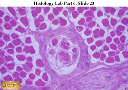 Muscle spindle -- a specialized sensory receptor for muscle stretch and position sense, as related particularly to unconscious maintenance of skeletal muscle tone and proper balance of postural muscle activity. The spindle is the encapsulated group of muscle fibers lying in the center of the field of regular skeletal muscle fibers, all cut in cross-section. The sensory nerve endings themselves (not visible here) wrap around the muscle fibers within the spindle. Such endings relay sensory information along dendrites within peripheral nerves, back to pseudounipolar cell bodies in a spinal ganglion, and thence to the spinal cord.
 Pacinian corpuscle -- another specialized sensory ending, this time for deep pressure. This particular view is from a whole mount of mesentery, so you are seeing the corpuscle three-dimensionally. They are also found in subcutaneous tissue, deep to skin. Notice the onion-like layers of specialized connective tissue surrounding a dark pink dendritic terminal. Again, the cell body for this dendrite lies in a spinal ganglion, and the axon of that same cell then proceeds into the spinal cord.
The following five slides show some specializations of the brain. First is an overview mid-sagittal cut of the brain, showing the many folds (or gyri) of the external cerebral cortex, and the much smaller, more delicate folds (or folia) of the cerebellar cortex seen to the left. As seen in this kind of cut, the cerebellar folia have a branching, tree-like appearance. (The brain stem is the solid-looking structure along the base of the brain, and continuous with the spinal cord at lower left.)
Section of cerebral cortex, showing cuts of two gyri. The pale cortex follows along the contours of the gyri. White matter (composed of nerve processes) lies below and stains a darker pink. Very little cytoarchitecture is seen with H&E stain.
Cerebral cortex stained with silver to show silhouettes of pyramidal cells. Now each triangular cell body can be seen, as well as the ascending apical dendrite, several basal dendrites, and a very fine descending axon. These are specialized multipolar neurons with such a definite shape that they can be recognized as such. You will learn more about them in Neuroscience.
Section of cerebellar cortex, showing several folia. Each folium has a central core of bright blue white matter, consisting of nerve processes entering and leaving the superficial cortex. The cortex has an external pale layer and a darker staining granular layer beneath it. Large Purkinje cells lie in a row between these two layers but are not visible at this magnification.
Higher magnification of cerebellar cortex, showing the row of large Purkinje cells lying between the outer and inner cortical layers. The stubs of the dendritic trees of the Purkinje cells look rather like "antlers" arising from the cell bodies. Very complex dendritic branchings actually extend throughout the molecular layer above the Purkinje c ells. A single axon leaves each Purkinje cell at its base and descends through the granular layer to deeper relay stations within the brain. Again, these are neurons with a very distinctive shape; you'll study their function and their connections next semester. |
| | | | ابراهيم الشنطي
Admin
عدد المساهمات : 70301
تاريخ التسجيل : 28/01/2013
العمر : 77
الموقع : الاردن
| | موضوع: رد: صور طبية هيستولوجى - Histology الإثنين 19 نوفمبر 2018, 12:28 am | |
| [size=48]Muscle[/size]
The cells of muscular tissue lie parallel to each other and, therefore, can be cut in either cross or longitudinal section and have to be distinguished from each other in both planes. They must also be distinguished from cuts of nerve and tendon. Watch for diagnostic features as you go along through these slides. [size=32]Longitudinal sections:[/size]Smooth muscle - long, slender central nuclei, lying within narrow, fusiform cells that lie parallel to each other in a smooth arrangement. (Muscle cells are often referred to as muscle fibers because of their narrowness and length.)
Smooth muscle - with cells more separated so as to see their extent and shape better, and the central position of their nuclei. A loose, irregular connective tissue (endomysium) lies between the cells. Nuclei seen in this c.t. belong to fibroblasts mainly.
Smooth muscle with wrinkled nuclei due to contraction of cells.
EM of smooth muscle showing typical "hairy" look of primarily filaments in the cytoplasm. Part of the cytoplasm is clear of filaments and shows mitochondria and polyribosomes. The cell membrane is at the lower right of the field and shows a few pinocytotic vesicles toward the extreme right. The left-hand extent of that same membrane seems darker and denser: probably a plaque, where filaments attach. The fuzzy density just outside the cell membrane is the basal lamina.
Skeletal muscle cells (fibers), with cross-striations and peripheral nuclei.
Higher power of skeletal muscle for details of cross-striations. Notice thin Z discs and heavy A bands. From one Z disc to the next is a sarcomere, the unit of muscle contraction. In the upper muscle cell notice shadowy myofibrils running longitudinally.
EM of several myofibrils running longitudinally through skeletal muscle cell. Between individual myofibrils lie the mitochondria (M) and glycogen (G) of the cytoplasm. Within each myofibril are the typical striations: A= A band; I= I band; Z= Z line; and H= H band. The banding is formed by the arrangement of myosin and actin filaments.
Cardiac muscle with cross-striations, dark intercalated discs, and centrally located nuclei. Notice too that the nuclei are stubby in appearance, and that they lie in a rather granular cytoplasm. Some of the intercalated discs form a straight line across muscle fibers; others make a step-like arrangement.
EM of intercalated disc between the ends of two cardiac muscle cells. Both desmosomes (1) and fasciae adheretes (2) are identified. Notice mitochondria and glycogen particles lying between myofibrils.
Another view of cardiac muscle showing wavy connective tissue (endomysium) between muscle cells. Also, notice capillaries with r.b.c.'s; muscle is a highly vascularized tissue. Some yellow granular cytoplasm can be seen inside the lower muscle cells, where myofibrils are parted. This picture also gives some indication of the branching of cardiac fibers.
This is a longitudinal section of peripheral nerve, for comparison with the three types of muscle. The foamy, pale look is due to the dissolving out of lipids from the myelin sheath. Note also the rounded constrictions of nodes of Ranvier.
Another comparison, this time with tendon (dense, regular, collagenous c.t.). Here you see very thin fibroblast nuclei compressed between collagen fibers and lined up in rows ("box-car").
Dense, fairly regular, collagenous tissue with mostly fibers and very few cells. Not as neatly arranged as the previous tissues.
[size=32]Cross-sections:[/size][size=32] [/size] [/size]Smooth muscle. Since the muscle cells are spindle-shaped, with tapered ends, the diameters of cross-cuts of individual cells vary considerably. Nuclei are central but appear only when the section goes through the widest part of the cell. Compare diameters of these cells with those in the next two slides, which are at the same magnification.
Cardiac muscle, with central nuclei surrounded by proportionally greater amounts of cytoplasm than previous smooth muscle. The "graininess" of the cytoplasm is due to cut ends of myofibrils. Remember that a very fine connective tissue endomysium lies between the individual muscle cells in all three types of muscle; often it is not well preserved because it collapses during fixation.
Skeletal muscle -- large, rounded cross-cuts of muscle cells, packed so full of myofibrils that nuclei are displaced to the periphery. (There is a capillary filled with pink rbc's in the upper middle field.)
A cross-cut of nerve for comparison. The pale central axons are surrounded by myelin sheaths that seem to have radiating lines in them due to the way the protein component of the sheath is preserved. All nuclei lie between nerve processes rather than in them.
A cross-cut of tendon to show fibroblasts compressed between thick pale collagenous fibers that they look stellate in shape. The cells look as if they are lying in "cracks" between the fibers; notice this on the right side of field particularly.
[size=32]Further details of muscle:[/size]
The inner surface of the heart showing large, pale-staining Purkinje fibers lying across the mid-portion of the picture. They are modified cardiac muscle fibers and seem mostly free of myofibrils except at the cell periphery, so that each cross-cut seems to have a darker pink rim and a pale center. The normal cardiac muscle fibers lie below in this micrograph and appear much smaller and more darkly stained than the Purkinje fibers.
Cross-cut of skeletal muscle to show connective tissue partitioning of muscle into groups or bundles of fibers. Endomysium is very delicate and lies between individual fibers, while perimysium is more visible and lies around a group of fibers. Epimysium is not seen here but ensheaths a whole muscle. In this picture notice the presence of small blood vessels in both perimysium and endomysium. Notice also the cross-cuts of myofibrils within the muscle cells, making them look grainy.
Longitudinal view of skeletal muscle cell with unusually clear cross-striations. This muscle is stretched, so that the A band is widely split.
a)Z disc
b)A band, split -- with pale H band in the middle
c)the line lies right in an H band
d)width of I band, with Z disc in the middle
e)pointing to a practically invisible thin line, the sarcolemma (or cell membrane), which lies outside the pale peripheral nucleus seen to the right
Diagram of contraction of skeletal muscle. On the left is the view with light microscopy. On the right are the thin actin filaments and thick myosin filaments seen in EM. Notice that the total width of the A band stays the same throughout and that the sliding in or out of the actin filaments determines the width of the H band. Consider which filaments you would see if you cut the muscle cross-wise through the I band, A band, or H band.
EM of cross-cut cardiac muscle showing thick myosin and thin actin filaments in a highly geometric arrangement.
Drawing of relationship (at EM level) of myofibrils to sarcoplasmic reticulum (smooth ER) and T-tubules in skeletal muscle. In this drawing the sarcoplasmic reticulum is labelled "sarcotubules" and "terminal cisternae". Notice that T-tubules are extensions of the sarcolemma (cell membrane, seen at right-hand edge), so that depolarization can spread along this part of the sarcolemma as well. (See diagrams and further explanation in your textbook.)
Same kind of diagram, this time for cardiac muscle. Note differences between the two in: [list="color: rgb(255, 238, 221); font-family: Arial, Tahoma, Helvetica, FreeSans, sans-serif; font-size: 13.524px; text-align: right; background-color: rgb(68, 21, 0);"] [*] their amount and arrangement of sarcoplasmic reticulum [*] the presence or near-absence of terminal cisterns (next to the T-tubules) [*] the position of T-tubules in relation to the A, I, and Z bands seen at the left. [/list] A triad consists of two terminal cisterns with a T-tubule in the middle. When the cisterns are not well developed, a true triad does not exist. A diad means two elements are together, as with one T-tubule and a neighboring bit of sarcoplasmic reticulum. NOTE: sarcoplasmic reticulum is just a form of smooth endoplasmic reticulum (SER). In muscle it is particularly associated with the release of calcium ions needed for contraction |
| | | | ابراهيم الشنطي
Admin
عدد المساهمات : 70301
تاريخ التسجيل : 28/01/2013
العمر : 77
الموقع : الاردن
| | موضوع: رد: صور طبية هيستولوجى - Histology الإثنين 19 نوفمبر 2018, 12:29 am | |
| Cardiovascular System
This capillary running through embryonic mesenchyme has a wall consisting solely of a single layer of endothelium. Notice that the lumen of the vessel is only slightly larger than the diameter of the r.b.c.'s within.
This sinusoid, like a capillary, has only an endothelial wall, but its lumen is characteristically considerably wider. Also, in some locations in the body (such as bone marrow, liver, and spleen) the endothelial cells of sinusoids are rather loosely joined together, thus permitting passage of blood cells between them.
In the middle of the field is a sinusoid (filled with orange-colored r.b.c.'s) in the marrow cavity of spongy bone. (The larger empty circles are fat cells.)
A capillary lying in the endomysium between skeletal muscle fibers. This one shows very dark endothelial nuclei and has 3 pink r.b.c.'s lined up in a row inside.
EM of different capillary endothelia.
- A) continuous, endothelium in capillary.
- B) fenestrated endothelium
- C) continuous, thin endothelium of lung capillary.
Note: basal lamina (1) under each one. Also pinocytotic vesicles (PV) and fenestrations (arrows).
Diagram of routes of transport across capillary or sinusoidal endothelial cells. (Notice that we now also consider discontinuous endothelium as well as the types you saw on the previous slide.)
- A) capillary lumen
- B) basal lamina
- C) pericapillary space (i.e., connective tissue ground substance)
- D) tight junction (fused outer leaflets)
- E) gap junction (2-4 nm or 20-40 A intercellular space)
- Fl-3) adjoining portions of separate endothelial cells that contact each other. Their ordinary intercellular distance is 150-200 A (15-20 nm).
"Spikes" on the outer leaflet of membrane = glycocalyx layer.
- 1 = diffusion through plasma membrane
- 2 = pinocytotic vesicles
- 3 = tight junction (impenetrable)
- 4 = gap junction (penetrable by small molecules)
- 5 = fenestration with closing membrane (flow allowed selectively)
- 6 = open fenestration within the cytoplasm of a given endothelial cell (easy passage of fluid and molecules)
- 7 = hole, discontinuity between endothelial cells (note that basal lamina is also gone)
Endothelium (simple squamous epithelium) lines the lumen of all blood and lymph vessels, as well as the heart. Here endothelial nuclei are seen ringing a venule (so identified because there is only a thin coat of connective tissue outside the endothelium, the lumen is too large to be a capillary, and because sinusoids don't occur in ordinary connective tissue areas like this.)
The smallest arterioles have a single layer of smooth muscle outside the endothelium, and a lumen hardly wider than a capillary. Toward the upper center of this field is a round, cross-cut arteriole with just one or two layers of muscle in the media. To the right is the more irregular, wider shape of a venule with only a thin adventitial wall.
EM of capillary. The inner nucleus and ring of cytoplasm is the endothelial cell around the lumen. The outer nucleus and ring of cytoplasm is a single pericyte wrapped around the vessel. Notice the thin gray line of basal lamina between the cytoplasm of the two cells and shared by both cells. There is also a basal lamina along the outer surface of the pericyte as well.
Small blood vessels, with 3-layered walls:
- a = small lymphatic with endothelial lining but no other organized wall.
- b = small vein (very thin muscle; pale c.t.).
- c = small artery (pink muscle; yellow c.t.).
These vessels are often found running together in the c.t. coats of body organs.
A small artery cut longitudinally. Notice the circular arrangement of smooth muscle cells cut tangentially at the left end. For most of the vessel, the muscle is cross-cut, looking almost like an epithelium. The real epithelium, however, is the simple squamous endothelium immediately lining the lumen, with thin, flat nuclei oriented longitudinally along the vessel.
A medium-sized (muscular) artery, showing the typical 3 wall layers:
- (1) tunica intima, consisting of endothelium lining the lumen, and a small amount of areolar c.t., a black and wavy inner elastic membrane divides this layer from the tunica media. (Only the elastic membrane shows up at this magnification.)
- (2) tunica media = smooth muscle (bluish color here) and some elastic fibers (black).
- (3) tunica adventitia = dense irregular c.t. (pink collagenic fibers, black elastic fibers). There is fat outside the adventitia.
Another medium-sized, muscular artery (also called a distributing artery). This is typical of the arteries you dissected in the arm; it usually runs with a vein and nerve. There is a characteristic inner elastic membrane (dark pink with arrow pointing to it) and a heavy circular muscle in tunica media. Note that a = adventitia.
EM photo of inner elastic membrane (white band). Endothelial cells are bunched up because the wall is contracted.
Medium-sized vein with a much less compact muscle layer than you saw in the preceding arteries. The tunica media is indicated by bar "a". Bar "b" = adventitia, which is at least as wide as the media, and often even wider. There is no evident inner elastic membrane. (Blood in the lumen stains red here.) To the right, compare sizes and walls of one small artery (d) and two very small veins (c) and (e).
Lymphatic vessel with a c.t. wall even thinner than a vein. There is a cut leaflet of a valve across the lumen. Material in the lumen contains no r.b.c.'s, mostly just structureless lymph and some lymphocytes. There are some fat cells and lymphocytes in the surrounding connective tissue.
Higher power of valve made of a core of fine c.t. with endothelium covering both surfaces. Valves in veins are constructed similarly.
Low power view of wall of aorta, an elastic artery:
- a = intima (endothelial lining plus thin layer of underlying c.t.).
- b = media (alternating layers of elastic membranes and smooth muscle, bound together by areolar c.t.) This is by far the thickest layer.
- c = adventitia (fairly dense c.t. carrying small blood vessels, the vasa vasorum
Detail of inner portion of aortic wall: "a" bar = depth of tunica intima (next to lumen). In the media there are many layers of wavy, dark-stained elastic membranes, alternating with the paler pink smooth muscle and areolar c.t. (Note that these are elastic membranes, not just fibers. Think of many layers of rubber sheets enwrapping the vessel, and you have cut across them and are looking at the cut edges. These sheets are fenestrated; i.e., they have holes in them, thus allowing passage of nutrients diffusing from the blood in the aortic lumen out into the tissues of the wall.) Arrows = nuclei of smooth muscle cells.
Detail of outer portion of aortic wall, showing blood vessels (vasa vasorum) in the adventitia. These vessels bring nutrients only to the outer one-third or so of the vessel wall.
- a = small nerve (very pale here).
- b = small vein - very little muscle, mostly pale c.t..
- c = small artery - considerable muscle, some c.t. (elastic fibers specifically stained and mixed with collagenic fibers). The c.t. adventitia just merges with the surrounding packing c.t..
Heart Inner surface of the heart, with pale, large Purkinje fibers lying in the subendocardial layer. Endocardium (or intima) is above. The beginning of the myocardium (media, cardiac muscle) is below.
 The heart wall, like blood vessels in general, has three main layers, though they are not called intima, media, and adventitia. As in vessels, however, the innermost and outermost layers are primarily connective tissue; the middle one is muscle --- in this case, cardiac muscle. From left to right, then, in this picture of ventricle wall, there is first a very thin endocardium, which consists primarily of an endothelial lining and a very small amount of connective tissue underneath it. The muscle layer, or myocardium is next and is by far the thickest layer and constitutes the bulk of the heart. To the far right is the epicardium, which contains considerable fat. In gross anatomy the epicardium is called the visceral layer of the serous pericardium; it has an outermost lining layer of mesothelium.
 A high magnification reminder of the appearance of cardiac muscle cut longitudinally, with central nucleus, branching fibers, and cross-striations. Muscle fibers spiral around the heart in all directions and can thus exert the necessary squeezing action as the heart contracts. Remember that these muscle cells are attached end to end by junctions at the intercalated disc. Axon terminals of autonomic neurons innervate some of the muscle cells, and the stimulus is spread to neighboring muscle cells by the intercalated discs and by gap junctions along the side walls of the cells.
Cardiac muscle in cross-section. Note also the many cross cut capillaries in the connective tissue endomysium between muscle fibers. As you might expect from the constant work the heart performs, it is a highly vascularized organ. Capillaries in this (or any) muscle have endothelium that is continuous and non-fenestrated.
The surface of the ventricular lumen is very irregular because of the presence of papillary muscles in the wall. These irregularities are, of course, lined with endothelium.
Low power of a Mallory-stained heart, showing two channels (above) that are continuous with the lumen of the left ventricle (below). The left-hand channel is the aorta, with some blue connective tissue in its wall. There is also one cusp of the semilunar valve, with its blue core of dense collagen. Remember that valves are lined over their entire surface by endothelium which is continuous with aortic endothelium above and the ventricular endothelium below. To the right in this picture is the atrioventricular channel, with chordae tendinae extending down from the mitral valve and attaching to the papillary muscles of the ventricle. Like valves, the chordae tendinae are also composed of dense collagenous connective tissue covered by an endothelial lining. |
| | | | ابراهيم الشنطي
Admin
عدد المساهمات : 70301
تاريخ التسجيل : 28/01/2013
العمر : 77
الموقع : الاردن
| | موضوع: رد: صور طبية هيستولوجى - Histology الإثنين 19 نوفمبر 2018, 12:30 am | |
| [size=32]Endocrine Glands[/size]
H & E section of hypophysis, with the darker pars distalis on the left and the paler pars nervosa on the right. The slit between the two parts is the remnant of Rathke's pouch. The very narrow band of darker-staining cells along the right-hand margin of the slit is all there is of the pars intermedia.
Sometimes the pars intermedia is cystic, as it is here. In this particular instance, the pars intermedia is fused with the pars distalis to the left, and the lumen of Rathke's pouch has disappeared. The split seen at the right is a fixation artifact; pars nervosa is at the lower right.
Pars distalis stained to show the 3 major types of secretory epithelial cells there:
- a = pale chromophobes
- b = blue basophil
- c = red acidophil
Notice that these cells are characteristically in cord-like clumps. The clumps are separated by a fine reticular fiber stroma (thin blue lines), where the blood capillaries lie. All endocrine glands are highly vascularized, since secretions go directly into capillaries (depending on the organ.) In endocrine glands the endothelium of these vessel s is typically continuous and fenestrated
Pars distalis stained with H & E. There are several purple basophils in the center and a good clump of pink acidophils above them. Most of the rest of the cells, rather pale ones, are the chromophobes.
Detail of the last picture with purple basophils toward the bottom center, pink acidophils at top center, and pale chromophobes scattered randomly. Very pale capillaries can be seen to the immediate left and right of the central clump of cells, lying within the delicate c.t. framework (stroma). Some secretion has collected in the center of the p icture. Be sure to consult your handout chart from class for hormones secreted and target organs affected.
Pale capillaries between clumps of acidophils of pars distalis. At the left, on the lower edge of the sinusoidal wall, is an elongate nucleus of an endothelial cell. EM would show that the endothelium here is continuous and fenestrated.
Two pale pink Herring bodies, collections of secretion in the pars nervosa of the pituitary. They represent accumulations of neurosecretion within the axons of neurons whose cell bodies lie in the hypothalamus of the brain.
Somatotroph from the anterior pituitary. Notice the numerous, spherical secretory granules.
Mammotroph from the anterior pituitary. The secretory granules are sparse and eliptical.
Low power view of thyroid gland with its characteristic colloid-filled follicles. This is the only endocrine gland that typically stores its hormonal secretion extracellularly before releasing it into the bloodstream.
Higher power of thyroid, showing follicular epithelium, which varies from simple squamous to simple cuboidal in height, depending on how distended the follicles are. Notice distended capillaries between follicles.
Another view of thyroid, showing a more consistently cuboidal epithelium. Lying between follicles, but not distinguishable here, are the C cells which secrete calcitonin. The arrows point to red capillaries; their endothelium is continuous and fenestrated.
EM of thyroid cell showing sparse microvilli on the apical surface that lies next to the stored colloid in the follicle.
Low power of parathyroid, showing random cords of epithelial cells which , on first glance at this power, might be mistaken for lymphocytes.
Another low power of parathyroid, showing a somewhat lobulated appearance and considerable adipose tissue intermingled with secretory portions.
Parathyroid at higher power, showing that the cells are actually tightly packed epithelial cords. The larger, pale pink cells in the middle and to the right are oxyphil cells; they have a smaller, darker nucleus and relatively larger amount of cytoplasm than the majority of cells, which are called chief cells (to the left in the photo). The chie f cells secrete parathormone; the significance of the oxyphil cells is not clear.
Panoramic view of adrenal gland, showing both cortex and medulla. The boundary between the two is about half way down from the surface. The zone glomerulosa is fairly dark and quite narrow; the zona fasciculata is the widest, palest layer of the cortex. The zone reticularis is darker and narrower. Medullary cells are larger and bluer in color here.
Higher power of adrenal cortex only. The zona glomerulosa at the top and the.zona reticularis at the bottom have more densely packed cells. The zona fasciculata in the middle has paler, larger cells; it is also the widest layer.
Adrenal cortex stained with Mallory-Azan. The c.t. capsule is dark blue. The zona glomerulosa (near the top) is narrow and has pale cells lying in loops and arches, with fine blue reticular fibers in between the cords. The zona fasciculata is quite wide and pale pinkish yellow. Cells in these layers are often pale and "foamy" looking because o f dissolved lipid of their steroid secretions. The zona reticularis shades from red to blue and merges indistinctly here with the medulla, which is also blue. The reticularis is wide here and extends nearly to the lower left corner of the field.
EM of adrenal cortical cell with cytoplasm filled with smooth endoplasmic reticulum, typical of steroid -secreting cells. Parts of two mitochondria are also visible here.
Detail of adrenal zona glomerulosa with large epithelial cells in looping and arching cords. Notice the blue areolar c.t. framework between the cords (mostly fine collagen fibers that would be classified as reticular).
Detail of adrenal zona fasciculata -- epithelial cells typically arranged in long, parallel columns. Notice the long endothelial nuclei of the sinusoids lying between the secretory cell cords. Endothelium here is generally considered to be fenestrated and continuous.
Junction between zona reticularis above (smaller cells in jumbled cords) and adrenal medulla below (larger cells). Blood in capillaries is red.
Detail of adrenal medullary cells in random epithelial cords.
Venous sinusoid in medulla of adrenal. The nucleus of an endothelial lining cell is seen in the center of the field. Remember that ovary, placenta, testis, kidney, GI tract and pancreas also have endocrine components! These organs are discussed separately as parts of other organ-systems. |
| | | | ابراهيم الشنطي
Admin
عدد المساهمات : 70301
تاريخ التسجيل : 28/01/2013
العمر : 77
الموقع : الاردن
| | موضوع: رد: صور طبية هيستولوجى - Histology الإثنين 19 نوفمبر 2018, 12:30 am | |
| [size=32]Respiratory Tract[/size]
Trachea, identified by the presence of hyaline cartilage in its wall. To the left of the cartilage is the mucosa, including epithelium and its underlying c.t.. The cartilage ring is immediately covered, on both surfaces, with bright pink perichondrium.
Trachea at higher magnification, showing pseudostratified ciliated columnar epithelium lining the lumen. A wide blood vessel lies in the lamina propria below. Hyaline cartilage is at the bottom of the picture.
Detail of pseudostratified ciliated columnar epithelium lining the trachea. Note the presence of pale goblet cells. Foreign particles are trapped in mucus secreted by goblet cells and then moved up (and out of) the respiratory tract by the beating cilia. A pale and quite thick basement membrane underlies this epithelium.
Posterior wall of the trachea with smooth muscle (pink strand near epithelium) replacing cartilage in the wall. The cartilage is basophilic in color. Structurally, it is a C-shaped ring, incomplete posteriorly, but with the trachealis muscle spanning the gap between the ends of the "C".
The trachealis muscle (smooth muscle) is the bright pink band in the middle of the field. Notice how cellular the c.t. under the epithelial layer is. Wandering blood cells, especially lymphocytes and eosinophils, are usually found here.
 Longitudinal, parasagittal section of trachea showing a series of darkly stained cartilage rings cut across. The rings are thicker anteriorly (at the right) than they are posteriorly (at the left). If this were a true mid-sagittal section, the cuts at the left would be of trachealis muscle instead of cartilage, so this section is clearly off-cen ter. The lining of the wide open lumen would be typical, pseudostratified "respiratory" epithelium. NOTE: Primary bronchi are essentially like the trachea in structure, and we have no example to show you. Lower portions of the respiratory tract appear in sections of lung and they show progressive loss of the various components characteristic of the trachea; that is, less and less cartilage, progressively lower epithelium, gradual loss of gob let cells, and finally loss of cilia and smooth muscle.
 An intrapulmonary bronchus with several separate pieces of hyaline cartilage in its wall. A thin layer of bright pink smooth muscle lies between the cartilage and the mucosa. (The mucosa, as always, consists of epithelium and underlying connective tissue.) In the upper left quadrant of the field is a large branch of the pulmonary artery, with it s bright pink tunica media (smooth muscle coat). Arterial branches accompany the branching respiratory tree all along the way.
An intrapulmonary bronchus with a patch of cartilage along its lower edge. Notice the field of alveoli and small branching terminals all around, typical of lung.
Detail of previous wall, showing the large chondrocytes of the cartilage. The epithelium lining the lumen looks pseudostratified still. A layer of pink smooth muscle lies between it and the cartilage. Considerable elastic tissue lies in the respiratory wall as well, but is not distinguishable in H&E stain.
A branching portion of the respiratory tree. No cartilage is visible here, so we'll say it's a bronchiole. Note a lymphatic nodule in the fork of the tree; such nodules may be found randomly along the respiratory tree. Note also the cross-cuts of pulmonary artery branches accompanying the tree. Cuts of alveoli of the lung fill the surrounding field.
Lung:
- a = alveoli, with thin interalveolar septa between them
- b = smooth muscle in its wall
- c = blood vessel, filled with r.b.c.'s
- d = bronchiole (again, no cartilage in its wall)
Lung:
- b = respiratory bronchiole with alveolus (a) in its wall. Most of the wall of the bronchiole has a definite line of dark along it, signifying a cuboidal or columnar epithelium (simple, rather than pseudostratified by now).
- d & c = alveolar duct. Its wall consists almost entirely of alveoli, which have only a simple squamous lining, too flat to be visible here.
- e = alveoli (the smallest respiratory units)
- f = blood vessel (branch of pulmonary artery still)
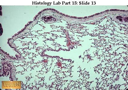 Another view of terminal branches of respiratory tree in the lung. In the middle of the field is a small terminal bronchiole branching off into three or four alveolar ducts to the right. The bronchiole has a definite, pink epithelial lining, while the walls of the alveolar ducts consist mainly of alveoli. The bronchiole is called terminal simpl y because it's the last generation of bronchiole before alveoli start to appear in the wall. Just before the alveolar ducts branch off, you can see a couple of small alveolar outpocketings in the bronchiole wall, thus making this short segment a respiratory bronchiole. (Note: along the top of this field is a wall of a bronchus, with two very sma ll, basophilic patches of cartilage just under the pink layer of smooth muscle.)
Lung
- a = inflated alveolar ducts
- c & d = blood vessels filled with r.b.c.'s
- e = bronchiole. Note pink simple columnar epithelium and absence of cartilage. Blue = connective tissue
The outer reaches of two lung lobules, with a connective tissue septum running vertically between them. The lower edge of the tissue here is the visceral pleura. Lymphatic vessels (a) and veins (b) run in the septa at the periphery of each lobule. Arteries, as we've noted previously, typically follow the branchings of the respiratory tree itsel f. At (c) there is a bronchiole. The mucosa lining its lumen is typically thrown into folds or scallops because of contraction of smooth muscle and elastic fibers in the wall.
Detail of inter-alveolar septa which form the shared walls of alveoli. The large spaces here are alveoli, filled with air. In the septa, orange is the red blood cells and brown is the nuclei of the capillary endothelium and alveolar cells. Every surface facing the alveolar air spaces is lined by simple squamous epithelium of the alveolar walls.
Detail of the septa between alveolar spaces. The small spaces within the septa are empty capillary lumens. Nuclei belong mainly to endothelial cells and alveolar epithelium; it would take EM to identify which is which with certainty. A few might also be fibroblasts or wandering c.t. cells. Very fine collagenous and elastic fibers also accompan y the capillaries. As you can see, especially in the capillaries in the center and in the upper left of the field, the combined thickness of capillary endothelium plus simple squamous alveolar epithelium separating blood from air, is extremely thin.
More detail of alveolar septa and walls. This time the capillary network is full of red blood cells.
EM showing basal lamina (1) between squamous alveolar epithelium (2 = Type I cell) and capillary endothelium (3). The nucleus at upper right belongs to the endothelial cell lining the capillary. The dark structure is a red blood cell. The capillary plus the alveolar linings on both sides constitute the interalveolar septum that lies between two alveolar spaces.
EM of alveolar wall with a Type II cell (or granular pneumocyte). It is a rounded or cuboidal cell, in contrast to the very flat Type I alveolar lining cell. To compare the differences in cell thickness, look in the lower part of the micrograph where you see capillary endothelium (1), basal lamina (2), and alveolar Type I epithelium (3) making up the very thin blood-air interface. The large, vacuolated, rather ragged looking vesicles in the cytoplasm of the Type II cell are lamellar bodies containing the precursor of alveolar surfactant. |
| | | | ابراهيم الشنطي
Admin
عدد المساهمات : 70301
تاريخ التسجيل : 28/01/2013
العمر : 77
الموقع : الاردن
| | موضوع: رد: صور طبية هيستولوجى - Histology الإثنين 19 نوفمبر 2018, 12:31 am | |
| [size=32]Urinary Tract - Kidney[/size]
Ureter, with typically stellate lumen. The transitional epithelium rests on a blue c.t. lamina propria. There is no boundary between lamina propria and the deeper c.t. submucosa. The muscularis externa stains a grayer blue in this slide; there are inner lonlitudinal and outer circular muscle layers visible here. On the outside is the blue c.t. adventitia with quite a bit of fat. (Mallory stain)
Ureter stained with H&E. Most layers of this wall are not well fixed, but the transitional epithelium and the stellate lumen are characteristic.
Transitional epithelium -- diagnostic for urinary tract. The surface cells are characteristically dome-shaped and puffy.
Transitional epithelium from a distended urinary bladder; compare with the contracted wall in the preceding slide. When stretched, the epithelium becomes very thin, with fewer layers of cells, and the surface cells tend to be flattened. There's only the one layer of flatter cells, however, which is quite different from the appearance of stratifi ed squamous epithelium (with which this might be confused.) See next slide.
Stratified squamous epithelium (non-keratinized) for comparison. Note that there are many layers of flattened cells, and, in fact, the whole epithelium is quite thick.
Anatomy of kidney. In the lower picture, notice positions of cortex, medullary pyramids, calyces, pelvis, blood vessels. Beginning with the minor calyces, and continuing on through the ureter, the lining is transitional epithelium.
Vascular arrangement. Follow renal artery to interlobar arteries to arcuate arteries to interlobular arteries. A kidney lobule lies between two interlobular arteries.
Papilla (right) projecting into calyx (left) lined with transitional epithelium.
Nephrons emptying into a collecting tubule. Notice that the closer to the medulla a glomerulus lies, the longer the loop of Henle is. Also notice that the inner zone of the medulla (lowest section of the picture) contains only thin limbs of the loop of Henle, plus collecting ducts. This is the area where the counter-current mechanism for urine concentration (carried out between the tubules and the surrounding peritubular capillaries) is most active. To make his drawing clear, the artist has made one accommodation that is not quite accurate histologically; namely, within the cortex, the straight positions of the nephrons (that is, the thick and thin portions of the loops of Henle) shoul d lie immediately next to the collecting duct, thus making up the medullary ray. The glomeruli and convoluted portions of the nephrons would then lie on either side of the ray. The ray is the central axis of a lobule.
 Cortex of kidney -- alternating straight rays and convoluted portions (with glomeruli). One longitudinally cut interlobular artery (pale pink contents) can be seen near the extreme left border of the picture, running up the middle of a convoluted portion, among glomeruli.
[*]a & b = interlobular vessels in cortex (vein larger than artery)
[*]Bar = the width of a straight (medullary) ray which constitutes the center of a lobule
[*]
Renal corpuscle with connection to proximal tubule at lower border. This, then, would be a cut through the urinary pole of the corpuscle.
Detail of renal corpuscle. Dark pink epithelium = proximal tubule. Lighter pink (as at upper top left) = distal tubule.
Dark pink = proximal tubule. Lighter, low cuboidal epithelium (as at top left) = distal tubule.
Proximal and distal convoluted tubules (EM). Distal has no brush border. Peritubular capillaries lie in the connective issue between tubules.
Higher EM of proximal tubule with its brush border (arrow), which indicates absorption by the cell.
EM of base of epithelium of proximal convoluted tubule. Note basement (basal) lamina and the great infolding of the cell membrane. These folds, plus the many mitochondria lying in them, tend to give the cytoplasm a striated look in light microscopy. The many folds also provide increased cell surface for passage of absorbed fluid and ions into t he peritubular capillary below.
Thin segment of Loop of Henle (in the middle), with a simple squamous lining.
Medullary region. There are some longitudinal cuts of pale collecting tubules (at left center) and several blood vessels filled with pale pink fluid (to the right). Epithelium of collecting tubules is regular, block-like, simple cuboidal, with unusually clear cell walls. Other tubules in the field are thick and thin limbs of loops of Henle.
Cross cuts in medulla. Two pale collecting tubules in the middle. Simple squamous lining indicates thin loops of Henle. Compare these with blood vessels, which contain r.b.c.'s. (Look for vessels up near top center and to right; also in lower left quadrant of field.) Notice the blue c.t. stroma in between the tubules.
Large pale tubules are collecting tubules, with clear epithelial cell boundaries. Brighter pink tubules are thick portions of loops of Henle; these are basically like distal convoluted tubules in their histology, so would be ascending limbs.
Renal corpuscle in Mallory stain to show bright orange r.b.c.'s in both the glomerular capillaries and the peritubular capillaries (among the convoluted tubules). Notice also the clear simple squamous epithelium of the parietal layer of Bowman's capsule outlining the whole renal corpuscle. The nuclei seen in the glomerulus could belong to endoth elium, podocytes, or mesangial cells.
Long thin artery leading to glomerulus (look in lower mid-picture). (Note long, thin endothelial nuclei lining the lumen. Circular muscle fibers have been cross-cut and look almost like a simple cuboidal epithelium outside the endothelium.)
Detail of wall of renal corpuscle. The space is the lumen of Bowman's capsule that receives glomerular filtrate from the capillary loops. Left wall is simple squamous parietal lining. The visceral lining of podocytes on the right wall of the space is too irregular to be seen clearly in light microscopy because it is following the curves of the individual capillaries.
EM of triangular shaped podocyte with its many terminal end feet (foot processes) touching the basement membrane (dark) which is shared on its other surface by endothelium of a capillary.
Detail of end feet of podocyte on the basement membrane. The basement membrane (basal lamina) is continuous, but the fenestrated capillary endothelium has pores. Glomerular filtrate passes from the capillary lumen, through the layers seen here, into the lumen of Bowman's capsule (where the foot processes are lying). Between the foot processes a re thin slit membranes.
EM of different capillary endothelia for further comparisons with other capillary walls.
- A) continuous, endothelium in capillary.
- B) fenestrated endothelium
- C) continuous, thin endothelium of lung capillary.
Note: basal lamina (1) under each one. Also pinocytotic vesicles (PV) and fenestrations (arrows).
Diagram of capillary loops in glomerulus and location of point of contact with distal convoluted tubule at base of loops. A histological section would show a closely packed portion of epithelial cells lining the distal tubule - the macula densa. Juxtaglomerular cells (J-G) would lie nearby in the wall of the afferent arteriole. The wedge-shaped space just above the distal tubule is called the polar cushion and would be filled with mesangial cells which are sometimes called Polkissen or Lacis cells in this location. Other mesangial cells extend up among the capillary loops of the glomerulus. By definition, the term.juxtaglomerular apparatus includes the J-G cells, macula densa, and pol ar cushion.
Diagram (top) of relation of juxtaglomerular (JG) cells to macula densa. Both areas sense the concentration of urine being produced, and by their action can alter it.
At lower right pole of glomerulus, note a triangular wedge of Polkissen cells just to the left of the straight row of macula densa cells. The latter are part of the epithelial wall of the distal tubule.
EM photo showing dark particles, which are secretory granules in the cytoplasm of juxtaglomerular cells. These cells are modified smooth muscle cells and secrete the hormone renin.
This slide, and the two that follow, show kidney tissue that has been imbedded in methacrylate and is unusually well preserved. This one shows the vascular pole of a renal corpuscle. The blood vessel is presumably an efferent arteriole because no "cuboidal" J-G cells are seen in its wall. (Note the biconcave shape of the rbc's in the vessel.) N otice also the clear parietal layer of Bowman's capsule. Surrounding the renal corpuscle are several cuts of the proximal tubule, with clear brush borders. There is one small distal tubule at bottom center of the field, slightly to the left.
This renal corpuscle has been sectioned through the urinary pole where the proximal tubule is continuous with the urinary space containing the urinary filtrate. Notice two cuts of distal tubule, one at left top edge of field, and one near right bottom edge.
A field of convoluted tubules, some with striated (brush) borders and some without. Notice that their cytoplasm often looks darkly striped or striated, from base to apex of the cells; this is because of all the mitochondria lined up between the basal infoldings of cell membrane. (Remember the EM you saw earlier in this set?) Notice also the small peritubular lying between the proximal and distal tubules; these branched off the efferent arteriole after it left the glomerulus; they will drain eventually into an interlobular vein. |
| | | | ابراهيم الشنطي
Admin
عدد المساهمات : 70301
تاريخ التسجيل : 28/01/2013
العمر : 77
الموقع : الاردن
| | موضوع: رد: صور طبية هيستولوجى - Histology الإثنين 19 نوفمبر 2018, 12:32 am | |
| [size=32]Gastrointestinal Tract[/size]
The digestive tract begins with the mouth, oral pharynx, and esophagus, all of which are lined by moist (non-keratinized) stratified squamous epithelium. In this diagram note the crossing of the digestive and respiratory tracts in the region of the pharynx. At that point there can be intermixed patches of pseudostratified columnar and stratified squamous epithelia.
The GI tract typically has a glandular mucosa (looks dark and thick here). Characteristics of the mucosa depend upon the function of the particular part of the tract concerned. This particular section is of colon, with lots of intestinal glands (crypts of Lieberkuhn) but no villi.
 Histological section of the GI tract, showing the basic layers clearly. The glandular mucosa (to the right) is quite dark because of all the epithelial and connective tissue nuclei it contains. A thin strip of pink marks the muscularis mucosae. Next comes the very dark pink submucosa which is mainly dense collagen fibers. Further left are the two, paler pink layers of the muscularis externa: a wide band of inner circular smooth muscle and a narrower band of outer longitudinal smooth muscle. Furthest left comes the serosa, so recognized because it has a "finished" edge of mesothelium. This is probably quite near the mesenteric attachment because there is so much adipose tissue and some fairly large blood vessels within the serosa.
Detail of mesothelium, looking down on its surface. The simple squamous cells fit together in a "pavement" effect. The intercellular boundaries have been silvered here.
Now we'll look at each part of the GI tract in turn, to see major identifying features. First is the esophagus showing the 4 major layers typical of GI wall, in order:
- Mucosa = stratified squamous epithelium on the surface with a paler connective tissue lamina propria underneath it and a darker, denser muscularis mucosae below the connective tissue.
- Submucosa = lighter connective tissue layer. Notice the submucosal gland with its duct extending up to the surface.
- Muscularis externa = darker inner circular and outer longitudinal smooth muscle bands. (Note presence of some brown is skeletal muscle intermixed.)
- Adventitia = very pale connective tissue at the bottom of the picture. Unlike a serosa, an adventitia just merges with surrounding connective tissue and has no "finished edge" of mesothelium
 Another section of esophagus, showing again the characteristic stratified squamous epithelium, the rather thick dense muscularis mucosae, and the very thick muscularis externa, with some patches of large-fibered skeletal muscle mixed with bands of smooth muscle. Notice that the muscle layers look more "solid" here than the lighter staining connec tive tissue layers. Blood vessels are evident in both the lamina propria and the submucosa. A piece of submucosal gland shows to the right.
Junction of esophagus (left) and cardiac stomach (right). There is a sudden change in epithelium, from stratified squamous to simple columnar. Mucosal glands of the cardiac portion of the stomach are simple coiled tubules, usually much shorter than pyloric glands, which tend to be longer and more crowded. (In our slides, you will see cardiac st omach only as a junction with esophagus, so there should be no confusion with pyloric stomach.)
Low power view of stomach through two folds in the wall. Dark pink represents mucosa, with many glands in it. Submucosa extends up into the folds. Note the thin pink line of the muscularis mucosae between the submucosa and the mucosal glands. The muscularis externa shows the usual two bands of muscle, is relatively thin, and has a definite oute r edge, indicating the presence of mesothelial serosa.
Gastric (fundic) glands of the mucosa of the main body of the stomach. They are typically straight and narrow, with shallow pits leading into long secretory portions. The surface epithelium dips down into pits and is simple columnar in type; these cells are all alike and secrete mucus. The chief and parietal cells of the lower portions of the gla nds will be discussed in detail in later slides. Notice the loose areolar lamina propria surrounding the glands, and the pink layer of muscularis mucosae at the base of the glands.
This section has sliced horizontally through several pits of gastric glands, showing the extremely regular simple columnar epithelial cells lining them. Lamina propria connective tissue lies between the pits.
Pyloric glands, which typically have long, narrow, tubular pits with a coiled glandular portion at the base. These glands produce a mucoid secretion. The thin muscularis mucosae and the thick muscularis externa are both solid looking and a deeper pink than the submucosa in between.
Mallory-stained pyloric mucosa, showing the coiled bases of the glands. There are no goblet cells in any portion of the stomach.
Transition from pylorus on left to duodenum on right. Notice the appearance of the duodenal glands of Brunner lying below the muscularis mucosae in the submucosa. These show up before duodenal villi appear on the surface.
Duodenum, showing villi and numerous short, darkly stained glands (crypts of Lieberkuhn) above the thin, pink muscularis mucosae, and pale glands of Brunner in the submucosa below. Peripheral to Brunner's glands are the solid-looking, pink, inner circular and outer longitudinal layers of the muscularis externa. The outermost edge is a "finished" one, so it is a serosa with mesothelium covering the surface; this means that the cut has been made on the duodenal surface lying next to the peritoneal cavity.
Detail of dark intestinal glands (crypts of Lieberkuhn) above and pale Brunner's alveolar units below the thin pink line of muscularis mucosae. Brunner's glands empty their mucous contents into the crypts. Their secretion is somewhat alkaline to neutralize the highly acid contents entering the duodenum from the stomach.
Another view of duodenum, with villi at the top, a band of straight, relatively short, narrow intestinal crypts below them, and then a whole field of pale, mucus-secreting Brunner's glands in the lower half of the picture (in the submucosa). Several tubules of Brunner's glands have pushed through the thin pink layer of muscularis mucosae, up into the mucosa; you can see one of them emptying into the base of a crypt on the right side of the picture. Through this route their secretions will reach the lumen of the gut. Notice the presence of goblet cells in the mucosal epithelium now that we've reached the small intestine.
Tangential cut through crypts; they are essentially small straight tubules lined with simple columnar epithelium. They secrete intestinal digestive enzymes and mucus. Notice the cellular connective tissue of the lamina propria lying between the crypts.
View of jejunum, showing long villi and short crypts. There are no longer any submucosal glands. The submucosa is thin and the muscularis externa is relatively thicker. There is a serosa because the jejunum is slung freely in mesentery.
Another view of small intestine to show how villi look when cut in cross-section. None of the villi are cut to show their full length.
Detail of cross-cut villi, showing goblet cells in the simple columnar absorptive epithelium. A cellular connective tissue lamina propria forms the core of each villus. More details will be seen a bit later.
Low power view of jejunum, in Mallory stain. Mucosa is purple; villi are quite tall, intestinal crypts short; notice the very thin purple line at the base of the mucosa (the muscularis mucosae). Submucosa is bright blue c.t. and extends up into the folds (plicae circulares). The two layers of the muscularis externa are purple-blue. There is a thin, light blue, c.t. serosa on the outside.
Wall of ileum, with villi and short crypts in the mucosa. There are now also Peyer's patches (dense lymphatic tissue which extends from the submucosa up into the mucosa, thus interrupting the thin, pink muscularis mucosae).
A reminder that an isolated lymphatic nodule can be found anywhere along the GI tract. This one is in the pyloric stomach. (Don't be fooled by what look like villi, making you think this is small intestine. Those are long pits going down to coiled glandular portions. Also, there are no goblet cells.)
Medium-power view of colon mucosa, with no villi and lots of crypts of Lieberkuhn. (Many bluish goblet cells are in the lining of the crypts.) The lamina propria around the crypts is heavily infiltrated with lymphocytes (typical of the colon).
Low power of the appendix, with its typically heavy concentration of lymphocytes in both the mucosa and submucosa. Crypts are still here but are less numerous than in the large and small intestines. Notice the presence of fat cells in the submucosa. The outermost coat also has an accumulation of fat.
Higher magnification of the same appendix, showing the heavy packing of lymphocytes in the wall. To the right, immediately beneath the crypts, you can see a narrow pink strip of muscularis mucosae, separating mucosa above from submucosa below. The dense diffuse distribution of lymphocytes indicates the presence of T-cells. A secondary lymphatic nodule with a germinal center lies to the left, indicating localized B-cell activity. Note the presence of goblet cells in the epithelium lining the surface and the crypts.
Detail of the appearance of crypts of Lieberkuhn as they appear in both the colon and appendix. The goblet cells are particularly numerous because of the need for lubrication of the gut contents. (The appendix, of course, is a vestigeal structure.) Notice also the numerous lymphocytes in the c.t. lamina propria.
 In the GI tract, the main differences from one part to the next are in the mucosa, and occasionally in the submucosa. The muscularis externa is pretty much the same in its basic pattern all along the way. Here you see the typical outermost layers, with a relatively thick inner circular band of smooth muscle and a usually thinner coat of outer lo nizitudinal smooth muscle. These directions are so consistent that you can determine the plane of section of the gut just by looking at the external muscle. In this particular case, the gut was cut in cross-section. This section also shows a serosa, with its "finished" edge of mesothelium. Any part of the gut that is slung in a mesentery has a serosa; any part that is retroperitoneal has an adventitia on the deep, buried surface and a serosa on the surface facing the peritoneal cavity. [Note: at the top of this structure you see an accumulation of lymphocytes in the submucosa.]
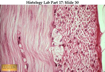 Another view of outer gut wall. At the extreme right there seems to be the beginning of some fat, so that is presumably the adventitia or serosa. This wall, then, like the previous one, has been cut transversely, as we can judge by the direction of cells in the musde layers. Between the two muscle layers is a pale, tangled-looking Auerbach's mye nteric p1exus. No cell bodies of neurons are visible, however.
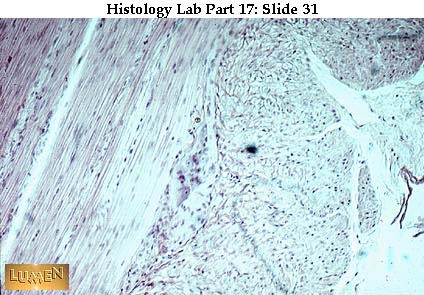 Autonomic (parasympathetic) ganglion cells lying between the two muscle layers of the muscularis externa (in middle of picture). There are two general locations where such ganglion cells are found in the gut: Meissner's (submucosal) plexus in the submucosa, and Auerbach's (myenteric) plexus between the two muscle layers. Both plexuses are parasy mpathetic, these being the postganglionic (or second) neurons in the basic two-cell autonomic chain. The preganglionic cells which synapse with most of the postganglionics of the gut lie in the dorsal motor nucleus of the vagus in the brain. (For the lowest portions of the gut, the preganglionics originate in the intermediate gray matter of the sacral spinal cord.)
Higher power of neuronal cell bodies of Auerbach's plexus in the middle of the picture. They are larger than surrounding cells and have the vesicular nuclei and dark prominent nucleoli typical of so many nerve cells.
In the lower left is a taenia coli, one of 3 strips of outer longitudinal muscle peculiar to this part of gut (the colon). Inner circular coat is continuous here above it, but the outer muscle coat is very thin at the right and thick at lower left where the taenia is. |
| | | | ابراهيم الشنطي
Admin
عدد المساهمات : 70301
تاريخ التسجيل : 28/01/2013
العمر : 77
الموقع : الاردن
| | موضوع: رد: صور طبية هيستولوجى - Histology الإثنين 19 نوفمبر 2018, 12:32 am | |
| [size=32]Gastrointestinal Tract[/size]
The digestive tract begins with the mouth, oral pharynx, and esophagus, all of which are lined by moist (non-keratinized) stratified squamous epithelium. In this diagram note the crossing of the digestive and respiratory tracts in the region of the pharynx. At that point there can be intermixed patches of pseudostratified columnar and stratified squamous epithelia.
The GI tract typically has a glandular mucosa (looks dark and thick here). Characteristics of the mucosa depend upon the function of the particular part of the tract concerned. This particular section is of colon, with lots of intestinal glands (crypts of Lieberkuhn) but no villi.
Histological section of the GI tract, showing the basic layers clearly. The glandular mucosa (to the right) is quite dark because of all the epithelial and connective tissue nuclei it contains. A thin strip of pink marks the muscularis mucosae. Next comes the very dark pink submucosa which is mainly dense collagen fibers. Further left are the two, paler pink layers of the muscularis externa: a wide band of inner circular smooth muscle and a narrower band of outer longitudinal smooth muscle. Furthest left comes the serosa, so recognized because it has a "finished" edge of mesothelium. This is probably quite near the mesenteric attachment because there is so much adipose tissue and some fairly large blood vessels within the serosa.
Detail of mesothelium, looking down on its surface. The simple squamous cells fit together in a "pavement" effect. The intercellular boundaries have been silvered here.
Now we'll look at each part of the GI tract in turn, to see major identifying features. First is the esophagus showing the 4 major layers typical of GI wall, in order:
- Mucosa = stratified squamous epithelium on the surface with a paler connective tissue lamina propria underneath it and a darker, denser muscularis mucosae below the connective tissue.
- Submucosa = lighter connective tissue layer. Notice the submucosal gland with its duct extending up to the surface.
- Muscularis externa = darker inner circular and outer longitudinal smooth muscle bands. (Note presence of some brown is skeletal muscle intermixed.)
- Adventitia = very pale connective tissue at the bottom of the picture. Unlike a serosa, an adventitia just merges with surrounding connective tissue and has no "finished edge" of mesothelium
Another section of esophagus, showing again the characteristic stratified squamous epithelium, the rather thick dense muscularis mucosae, and the very thick muscularis externa, with some patches of large-fibered skeletal muscle mixed with bands of smooth muscle. Notice that the muscle layers look more "solid" here than the lighter staining connec tive tissue layers. Blood vessels are evident in both the lamina propria and the submucosa. A piece of submucosal gland shows to the right.
Junction of esophagus (left) and cardiac stomach (right). There is a sudden change in epithelium, from stratified squamous to simple columnar. Mucosal glands of the cardiac portion of the stomach are simple coiled tubules, usually much shorter than pyloric glands, which tend to be longer and more crowded. (In our slides, you will see cardiac st omach only as a junction with esophagus, so there should be no confusion with pyloric stomach.)
Low power view of stomach through two folds in the wall. Dark pink represents mucosa, with many glands in it. Submucosa extends up into the folds. Note the thin pink line of the muscularis mucosae between the submucosa and the mucosal glands. The muscularis externa shows the usual two bands of muscle, is relatively thin, and has a definite oute r edge, indicating the presence of mesothelial serosa.
Gastric (fundic) glands of the mucosa of the main body of the stomach. They are typically straight and narrow, with shallow pits leading into long secretory portions. The surface epithelium dips down into pits and is simple columnar in type; these cells are all alike and secrete mucus. The chief and parietal cells of the lower portions of the gla nds will be discussed in detail in later slides. Notice the loose areolar lamina propria surrounding the glands, and the pink layer of muscularis mucosae at the base of the glands.
This section has sliced horizontally through several pits of gastric glands, showing the extremely regular simple columnar epithelial cells lining them. Lamina propria connective tissue lies between the pits.
Pyloric glands, which typically have long, narrow, tubular pits with a coiled glandular portion at the base. These glands produce a mucoid secretion. The thin muscularis mucosae and the thick muscularis externa are both solid looking and a deeper pink than the submucosa in between.
Mallory-stained pyloric mucosa, showing the coiled bases of the glands. There are no goblet cells in any portion of the stomach.
Transition from pylorus on left to duodenum on right. Notice the appearance of the duodenal glands of Brunner lying below the muscularis mucosae in the submucosa. These show up before duodenal villi appear on the surface.
Duodenum, showing villi and numerous short, darkly stained glands (crypts of Lieberkuhn) above the thin, pink muscularis mucosae, and pale glands of Brunner in the submucosa below. Peripheral to Brunner's glands are the solid-looking, pink, inner circular and outer longitudinal layers of the muscularis externa. The outermost edge is a "finished" one, so it is a serosa with mesothelium covering the surface; this means that the cut has been made on the duodenal surface lying next to the peritoneal cavity.
Detail of dark intestinal glands (crypts of Lieberkuhn) above and pale Brunner's alveolar units below the thin pink line of muscularis mucosae. Brunner's glands empty their mucous contents into the crypts. Their secretion is somewhat alkaline to neutralize the highly acid contents entering the duodenum from the stomach.
Another view of duodenum, with villi at the top, a band of straight, relatively short, narrow intestinal crypts below them, and then a whole field of pale, mucus-secreting Brunner's glands in the lower half of the picture (in the submucosa). Several tubules of Brunner's glands have pushed through the thin pink layer of muscularis mucosae, up into the mucosa; you can see one of them emptying into the base of a crypt on the right side of the picture. Through this route their secretions will reach the lumen of the gut. Notice the presence of goblet cells in the mucosal epithelium now that we've reached the small intestine.
Tangential cut through crypts; they are essentially small straight tubules lined with simple columnar epithelium. They secrete intestinal digestive enzymes and mucus. Notice the cellular connective tissue of the lamina propria lying between the crypts.
View of jejunum, showing long villi and short crypts. There are no longer any submucosal glands. The submucosa is thin and the muscularis externa is relatively thicker. There is a serosa because the jejunum is slung freely in mesentery.
Another view of small intestine to show how villi look when cut in cross-section. None of the villi are cut to show their full length.
Detail of cross-cut villi, showing goblet cells in the simple columnar absorptive epithelium. A cellular connective tissue lamina propria forms the core of each villus. More details will be seen a bit later.
Low power view of jejunum, in Mallory stain. Mucosa is purple; villi are quite tall, intestinal crypts short; notice the very thin purple line at the base of the mucosa (the muscularis mucosae). Submucosa is bright blue c.t. and extends up into the folds (plicae circulares). The two layers of the muscularis externa are purple-blue. There is a thin, light blue, c.t. serosa on the outside.
Wall of ileum, with villi and short crypts in the mucosa. There are now also Peyer's patches (dense lymphatic tissue which extends from the submucosa up into the mucosa, thus interrupting the thin, pink muscularis mucosae).
A reminder that an isolated lymphatic nodule can be found anywhere along the GI tract. This one is in the pyloric stomach. (Don't be fooled by what look like villi, making you think this is small intestine. Those are long pits going down to coiled glandular portions. Also, there are no goblet cells.)
Medium-power view of colon mucosa, with no villi and lots of crypts of Lieberkuhn. (Many bluish goblet cells are in the lining of the crypts.) The lamina propria around the crypts is heavily infiltrated with lymphocytes (typical of the colon).
Low power of the appendix, with its typically heavy concentration of lymphocytes in both the mucosa and submucosa. Crypts are still here but are less numerous than in the large and small intestines. Notice the presence of fat cells in the submucosa. The outermost coat also has an accumulation of fat.
Higher magnification of the same appendix, showing the heavy packing of lymphocytes in the wall. To the right, immediately beneath the crypts, you can see a narrow pink strip of muscularis mucosae, separating mucosa above from submucosa below. The dense diffuse distribution of lymphocytes indicates the presence of T-cells. A secondary lymphatic nodule with a germinal center lies to the left, indicating localized B-cell activity. Note the presence of goblet cells in the epithelium lining the surface and the crypts.
Detail of the appearance of crypts of Lieberkuhn as they appear in both the colon and appendix. The goblet cells are particularly numerous because of the need for lubrication of the gut contents. (The appendix, of course, is a vestigeal structure.) Notice also the numerous lymphocytes in the c.t. lamina propria.
In the GI tract, the main differences from one part to the next are in the mucosa, and occasionally in the submucosa. The muscularis externa is pretty much the same in its basic pattern all along the way. Here you see the typical outermost layers, with a relatively thick inner circular band of smooth muscle and a usually thinner coat of outer lo nizitudinal smooth muscle. These directions are so consistent that you can determine the plane of section of the gut just by looking at the external muscle. In this particular case, the gut was cut in cross-section. This section also shows a serosa, with its "finished" edge of mesothelium. Any part of the gut that is slung in a mesentery has a serosa; any part that is retroperitoneal has an adventitia on the deep, buried surface and a serosa on the surface facing the peritoneal cavity. [Note: at the top of this structure you see an accumulation of lymphocytes in the submucosa.]
Another view of outer gut wall. At the extreme right there seems to be the beginning of some fat, so that is presumably the adventitia or serosa. This wall, then, like the previous one, has been cut transversely, as we can judge by the direction of cells in the musde layers. Between the two muscle layers is a pale, tangled-looking Auerbach's mye nteric p1exus. No cell bodies of neurons are visible, however.
Autonomic (parasympathetic) ganglion cells lying between the two muscle layers of the muscularis externa (in middle of picture). There are two general locations where such ganglion cells are found in the gut: Meissner's (submucosal) plexus in the submucosa, and Auerbach's (myenteric) plexus between the two muscle layers. Both plexuses are parasy mpathetic, these being the postganglionic (or second) neurons in the basic two-cell autonomic chain. The preganglionic cells which synapse with most of the postganglionics of the gut lie in the dorsal motor nucleus of the vagus in the brain. (For the lowest portions of the gut, the preganglionics originate in the intermediate gray matter of the sacral spinal cord.)
Higher power of neuronal cell bodies of Auerbach's plexus in the middle of the picture. They are larger than surrounding cells and have the vesicular nuclei and dark prominent nucleoli typical of so many nerve cells.
In the lower left is a taenia coli, one of 3 strips of outer longitudinal muscle peculiar to this part of gut (the colon). Inner circular coat is continuous here above it, but the outer muscle coat is very thin at the right and thick at lower left where the taenia is. |
| | | | ابراهيم الشنطي
Admin
عدد المساهمات : 70301
تاريخ التسجيل : 28/01/2013
العمر : 77
الموقع : الاردن
| | موضوع: رد: صور طبية هيستولوجى - Histology الإثنين 19 نوفمبر 2018, 12:33 am | |
| [size=32]Endocrine Glands[/size]
H & E section of hypophysis, with the darker pars distalis on the left and the paler pars nervosa on the right. The slit between the two parts is the remnant of Rathke's pouch. The very narrow band of darker-staining cells along the right-hand margin of the slit is all there is of the pars intermedia.
Sometimes the pars intermedia is cystic, as it is here. In this particular instance, the pars intermedia is fused with the pars distalis to the left, and the lumen of Rathke's pouch has disappeared. The split seen at the right is a fixation artifact; pars nervosa is at the lower right.
Pars distalis stained to show the 3 major types of secretory epithelial cells there:
- a = pale chromophobes
- b = blue basophil
- c = red acidophil
Notice that these cells are characteristically in cord-like clumps. The clumps are separated by a fine reticular fiber stroma (thin blue lines), where the blood capillaries lie. All endocrine glands are highly vascularized, since secretions go directly into capillaries (depending on the organ.) In endocrine glands the endothelium of these vessel s is typically continuous and fenestrated
Pars distalis stained with H & E. There are several purple basophils in the center and a good clump of pink acidophils above them. Most of the rest of the cells, rather pale ones, are the chromophobes.
Detail of the last picture with purple basophils toward the bottom center, pink acidophils at top center, and pale chromophobes scattered randomly. Very pale capillaries can be seen to the immediate left and right of the central clump of cells, lying within the delicate c.t. framework (stroma). Some secretion has collected in the center of the p icture. Be sure to consult your handout chart from class for hormones secreted and target organs affected.
Pale capillaries between clumps of acidophils of pars distalis. At the left, on the lower edge of the sinusoidal wall, is an elongate nucleus of an endothelial cell. EM would show that the endothelium here is continuous and fenestrated.
Two pale pink Herring bodies, collections of secretion in the pars nervosa of the pituitary. They represent accumulations of neurosecretion within the axons of neurons whose cell bodies lie in the hypothalamus of the brain.
Somatotroph from the anterior pituitary. Notice the numerous, spherical secretory granules.
Mammotroph from the anterior pituitary. The secretory granules are sparse and eliptical.
Low power view of thyroid gland with its characteristic colloid-filled follicles. This is the only endocrine gland that typically stores its hormonal secretion extracellularly before releasing it into the bloodstream.
Higher power of thyroid, showing follicular epithelium, which varies from simple squamous to simple cuboidal in height, depending on how distended the follicles are. Notice distended capillaries between follicles.
Another view of thyroid, showing a more consistently cuboidal epithelium. Lying between follicles, but not distinguishable here, are the C cells which secrete calcitonin. The arrows point to red capillaries; their endothelium is continuous and fenestrated.
EM of thyroid cell showing sparse microvilli on the apical surface that lies next to the stored colloid in the follicle.
Low power of parathyroid, showing random cords of epithelial cells which , on first glance at this power, might be mistaken for lymphocytes.
Another low power of parathyroid, showing a somewhat lobulated appearance and considerable adipose tissue intermingled with secretory portions.
Parathyroid at higher power, showing that the cells are actually tightly packed epithelial cords. The larger, pale pink cells in the middle and to the right are oxyphil cells; they have a smaller, darker nucleus and relatively larger amount of cytoplasm than the majority of cells, which are called chief cells (to the left in the photo). The chie f cells secrete parathormone; the significance of the oxyphil cells is not clear.
Panoramic view of adrenal gland, showing both cortex and medulla. The boundary between the two is about half way down from the surface. The zone glomerulosa is fairly dark and quite narrow; the zona fasciculata is the widest, palest layer of the cortex. The zone reticularis is darker and narrower. Medullary cells are larger and bluer in color here.
Higher power of adrenal cortex only. The zona glomerulosa at the top and the.zona reticularis at the bottom have more densely packed cells. The zona fasciculata in the middle has paler, larger cells; it is also the widest layer.
Adrenal cortex stained with Mallory-Azan. The c.t. capsule is dark blue. The zona glomerulosa (near the top) is narrow and has pale cells lying in loops and arches, with fine blue reticular fibers in between the cords. The zona fasciculata is quite wide and pale pinkish yellow. Cells in these layers are often pale and "foamy" looking because o f dissolved lipid of their steroid secretions. The zona reticularis shades from red to blue and merges indistinctly here with the medulla, which is also blue. The reticularis is wide here and extends nearly to the lower left corner of the field.
EM of adrenal cortical cell with cytoplasm filled with smooth endoplasmic reticulum, typical of steroid -secreting cells. Parts of two mitochondria are also visible here.
Detail of adrenal zona glomerulosa with large epithelial cells in looping and arching cords. Notice the blue areolar c.t. framework between the cords (mostly fine collagen fibers that would be classified as reticular).
Detail of adrenal zona fasciculata -- epithelial cells typically arranged in long, parallel columns. Notice the long endothelial nuclei of the sinusoids lying between the secretory cell cords. Endothelium here is generally considered to be fenestrated and continuous.
Junction between zona reticularis above (smaller cells in jumbled cords) and adrenal medulla below (larger cells). Blood in capillaries is red.
Detail of adrenal medullary cells in random epithelial cords.
Venous sinusoid in medulla of adrenal. The nucleus of an endothelial lining cell is seen in the center of the field. Remember that ovary, placenta, testis, kidney, GI tract and pancreas also have endocrine components! These organs are discussed separately as parts of other organ-systems. |
| | | | ابراهيم الشنطي
Admin
عدد المساهمات : 70301
تاريخ التسجيل : 28/01/2013
العمر : 77
الموقع : الاردن
| | موضوع: رد: صور طبية هيستولوجى - Histology الإثنين 19 نوفمبر 2018, 12:33 am | |
| [size=32]Respiratory Tract[/size]
Trachea, identified by the presence of hyaline cartilage in its wall. To the left of the cartilage is the mucosa, including epithelium and its underlying c.t.. The cartilage ring is immediately covered, on both surfaces, with bright pink perichondrium.
Trachea at higher magnification, showing pseudostratified ciliated columnar epithelium lining the lumen. A wide blood vessel lies in the lamina propria below. Hyaline cartilage is at the bottom of the picture.
Detail of pseudostratified ciliated columnar epithelium lining the trachea. Note the presence of pale goblet cells. Foreign particles are trapped in mucus secreted by goblet cells and then moved up (and out of) the respiratory tract by the beating cilia. A pale and quite thick basement membrane underlies this epithelium.
Posterior wall of the trachea with smooth muscle (pink strand near epithelium) replacing cartilage in the wall. The cartilage is basophilic in color. Structurally, it is a C-shaped ring, incomplete posteriorly, but with the trachealis muscle spanning the gap between the ends of the "C".
The trachealis muscle (smooth muscle) is the bright pink band in the middle of the field. Notice how cellular the c.t. under the epithelial layer is. Wandering blood cells, especially lymphocytes and eosinophils, are usually found here.
Longitudinal, parasagittal section of trachea showing a series of darkly stained cartilage rings cut across. The rings are thicker anteriorly (at the right) than they are posteriorly (at the left). If this were a true mid-sagittal section, the cuts at the left would be of trachealis muscle instead of cartilage, so this section is clearly off-cen ter. The lining of the wide open lumen would be typical, pseudostratified "respiratory" epithelium. NOTE: Primary bronchi are essentially like the trachea in structure, and we have no example to show you. Lower portions of the respiratory tract appear in sections of lung and they show progressive loss of the various components characteristic of the trachea; that is, less and less cartilage, progressively lower epithelium, gradual loss of gob let cells, and finally loss of cilia and smooth muscle.
An intrapulmonary bronchus with several separate pieces of hyaline cartilage in its wall. A thin layer of bright pink smooth muscle lies between the cartilage and the mucosa. (The mucosa, as always, consists of epithelium and underlying connective tissue.) In the upper left quadrant of the field is a large branch of the pulmonary artery, with it s bright pink tunica media (smooth muscle coat). Arterial branches accompany the branching respiratory tree all along the way.
An intrapulmonary bronchus with a patch of cartilage along its lower edge. Notice the field of alveoli and small branching terminals all around, typical of lung.
Detail of previous wall, showing the large chondrocytes of the cartilage. The epithelium lining the lumen looks pseudostratified still. A layer of pink smooth muscle lies between it and the cartilage. Considerable elastic tissue lies in the respiratory wall as well, but is not distinguishable in H&E stain.
A branching portion of the respiratory tree. No cartilage is visible here, so we'll say it's a bronchiole. Note a lymphatic nodule in the fork of the tree; such nodules may be found randomly along the respiratory tree. Note also the cross-cuts of pulmonary artery branches accompanying the tree. Cuts of alveoli of the lung fill the surrounding field.
Lung:
- a = alveoli, with thin interalveolar septa between them
- b = smooth muscle in its wall
- c = blood vessel, filled with r.b.c.'s
- d = bronchiole (again, no cartilage in its wall)
Lung:
- b = respiratory bronchiole with alveolus (a) in its wall. Most of the wall of the bronchiole has a definite line of dark along it, signifying a cuboidal or columnar epithelium (simple, rather than pseudostratified by now).
- d & c = alveolar duct. Its wall consists almost entirely of alveoli, which have only a simple squamous lining, too flat to be visible here.
- e = alveoli (the smallest respiratory units)
- f = blood vessel (branch of pulmonary artery still)
Another view of terminal branches of respiratory tree in the lung. In the middle of the field is a small terminal bronchiole branching off into three or four alveolar ducts to the right. The bronchiole has a definite, pink epithelial lining, while the walls of the alveolar ducts consist mainly of alveoli. The bronchiole is called terminal simpl y because it's the last generation of bronchiole before alveoli start to appear in the wall. Just before the alveolar ducts branch off, you can see a couple of small alveolar outpocketings in the bronchiole wall, thus making this short segment a respiratory bronchiole. (Note: along the top of this field is a wall of a bronchus, with two very sma ll, basophilic patches of cartilage just under the pink layer of smooth muscle.)
Lung
- a = inflated alveolar ducts
- c & d = blood vessels filled with r.b.c.'s
- e = bronchiole. Note pink simple columnar epithelium and absence of cartilage. Blue = connective tissue
The outer reaches of two lung lobules, with a connective tissue septum running vertically between them. The lower edge of the tissue here is the visceral pleura. Lymphatic vessels (a) and veins (b) run in the septa at the periphery of each lobule. Arteries, as we've noted previously, typically follow the branchings of the respiratory tree itsel f. At (c) there is a bronchiole. The mucosa lining its lumen is typically thrown into folds or scallops because of contraction of smooth muscle and elastic fibers in the wall.
Detail of inter-alveolar septa which form the shared walls of alveoli. The large spaces here are alveoli, filled with air. In the septa, orange is the red blood cells and brown is the nuclei of the capillary endothelium and alveolar cells. Every surface facing the alveolar air spaces is lined by simple squamous epithelium of the alveolar walls.
Detail of the septa between alveolar spaces. The small spaces within the septa are empty capillary lumens. Nuclei belong mainly to endothelial cells and alveolar epithelium; it would take EM to identify which is which with certainty. A few might also be fibroblasts or wandering c.t. cells. Very fine collagenous and elastic fibers also accompan y the capillaries. As you can see, especially in the capillaries in the center and in the upper left of the field, the combined thickness of capillary endothelium plus simple squamous alveolar epithelium separating blood from air, is extremely thin.
More detail of alveolar septa and walls. This time the capillary network is full of red blood cells.
EM showing basal lamina (1) between squamous alveolar epithelium (2 = Type I cell) and capillary endothelium (3). The nucleus at upper right belongs to the endothelial cell lining the capillary. The dark structure is a red blood cell. The capillary plus the alveolar linings on both sides constitute the interalveolar septum that lies between two alveolar spaces.
EM of alveolar wall with a Type II cell (or granular pneumocyte). It is a rounded or cuboidal cell, in contrast to the very flat Type I alveolar lining cell. To compare the differences in cell thickness, look in the lower part of the micrograph where you see capillary endothelium (1), basal lamina (2), and alveolar Type I epithelium (3) making up the very thin blood-air interface. The large, vacuolated, rather ragged looking vesicles in the cytoplasm of the Type II cell are lamellar bodies containing the precursor of alveolar surfactant. |
| | | | ابراهيم الشنطي
Admin
عدد المساهمات : 70301
تاريخ التسجيل : 28/01/2013
العمر : 77
الموقع : الاردن
| | موضوع: رد: صور طبية هيستولوجى - Histology الإثنين 19 نوفمبر 2018, 12:35 am | |
| [size=32]Gastrointestinal Tract[/size]
The digestive tract begins with the mouth, oral pharynx, and esophagus, all of which are lined by moist (non-keratinized) stratified squamous epithelium. In this diagram note the crossing of the digestive and respiratory tracts in the region of the pharynx. At that point there can be intermixed patches of pseudostratified columnar and stratified squamous epithelia.
The GI tract typically has a glandular mucosa (looks dark and thick here). Characteristics of the mucosa depend upon the function of the particular part of the tract concerned. This particular section is of colon, with lots of intestinal glands (crypts of Lieberkuhn) but no villi.
Histological section of the GI tract, showing the basic layers clearly. The glandular mucosa (to the right) is quite dark because of all the epithelial and connective tissue nuclei it contains. A thin strip of pink marks the muscularis mucosae. Next comes the very dark pink submucosa which is mainly dense collagen fibers. Further left are the two, paler pink layers of the muscularis externa: a wide band of inner circular smooth muscle and a narrower band of outer longitudinal smooth muscle. Furthest left comes the serosa, so recognized because it has a "finished" edge of mesothelium. This is probably quite near the mesenteric attachment because there is so much adipose tissue and some fairly large blood vessels within the serosa.
Detail of mesothelium, looking down on its surface. The simple squamous cells fit together in a "pavement" effect. The intercellular boundaries have been silvered here.
Now we'll look at each part of the GI tract in turn, to see major identifying features. First is the esophagus showing the 4 major layers typical of GI wall, in order:
- Mucosa = stratified squamous epithelium on the surface with a paler connective tissue lamina propria underneath it and a darker, denser muscularis mucosae below the connective tissue.
- Submucosa = lighter connective tissue layer. Notice the submucosal gland with its duct extending up to the surface.
- Muscularis externa = darker inner circular and outer longitudinal smooth muscle bands. (Note presence of some brown is skeletal muscle intermixed.)
- Adventitia = very pale connective tissue at the bottom of the picture. Unlike a serosa, an adventitia just merges with surrounding connective tissue and has no "finished edge" of mesothelium
Another section of esophagus, showing again the characteristic stratified squamous epithelium, the rather thick dense muscularis mucosae, and the very thick muscularis externa, with some patches of large-fibered skeletal muscle mixed with bands of smooth muscle. Notice that the muscle layers look more "solid" here than the lighter staining connec tive tissue layers. Blood vessels are evident in both the lamina propria and the submucosa. A piece of submucosal gland shows to the right.
Junction of esophagus (left) and cardiac stomach (right). There is a sudden change in epithelium, from stratified squamous to simple columnar. Mucosal glands of the cardiac portion of the stomach are simple coiled tubules, usually much shorter than pyloric glands, which tend to be longer and more crowded. (In our slides, you will see cardiac st omach only as a junction with esophagus, so there should be no confusion with pyloric stomach.)
Low power view of stomach through two folds in the wall. Dark pink represents mucosa, with many glands in it. Submucosa extends up into the folds. Note the thin pink line of the muscularis mucosae between the submucosa and the mucosal glands. The muscularis externa shows the usual two bands of muscle, is relatively thin, and has a definite oute r edge, indicating the presence of mesothelial serosa.
Gastric (fundic) glands of the mucosa of the main body of the stomach. They are typically straight and narrow, with shallow pits leading into long secretory portions. The surface epithelium dips down into pits and is simple columnar in type; these cells are all alike and secrete mucus. The chief and parietal cells of the lower portions of the gla nds will be discussed in detail in later slides. Notice the loose areolar lamina propria surrounding the glands, and the pink layer of muscularis mucosae at the base of the glands.
This section has sliced horizontally through several pits of gastric glands, showing the extremely regular simple columnar epithelial cells lining them. Lamina propria connective tissue lies between the pits.
Pyloric glands, which typically have long, narrow, tubular pits with a coiled glandular portion at the base. These glands produce a mucoid secretion. The thin muscularis mucosae and the thick muscularis externa are both solid looking and a deeper pink than the submucosa in between.
Mallory-stained pyloric mucosa, showing the coiled bases of the glands. There are no goblet cells in any portion of the stomach.
Transition from pylorus on left to duodenum on right. Notice the appearance of the duodenal glands of Brunner lying below the muscularis mucosae in the submucosa. These show up before duodenal villi appear on the surface.
Duodenum, showing villi and numerous short, darkly stained glands (crypts of Lieberkuhn) above the thin, pink muscularis mucosae, and pale glands of Brunner in the submucosa below. Peripheral to Brunner's glands are the solid-looking, pink, inner circular and outer longitudinal layers of the muscularis externa. The outermost edge is a "finished" one, so it is a serosa with mesothelium covering the surface; this means that the cut has been made on the duodenal surface lying next to the peritoneal cavity.
Detail of dark intestinal glands (crypts of Lieberkuhn) above and pale Brunner's alveolar units below the thin pink line of muscularis mucosae. Brunner's glands empty their mucous contents into the crypts. Their secretion is somewhat alkaline to neutralize the highly acid contents entering the duodenum from the stomach.
Another view of duodenum, with villi at the top, a band of straight, relatively short, narrow intestinal crypts below them, and then a whole field of pale, mucus-secreting Brunner's glands in the lower half of the picture (in the submucosa). Several tubules of Brunner's glands have pushed through the thin pink layer of muscularis mucosae, up into the mucosa; you can see one of them emptying into the base of a crypt on the right side of the picture. Through this route their secretions will reach the lumen of the gut. Notice the presence of goblet cells in the mucosal epithelium now that we've reached the small intestine.
Tangential cut through crypts; they are essentially small straight tubules lined with simple columnar epithelium. They secrete intestinal digestive enzymes and mucus. Notice the cellular connective tissue of the lamina propria lying between the crypts.
View of jejunum, showing long villi and short crypts. There are no longer any submucosal glands. The submucosa is thin and the muscularis externa is relatively thicker. There is a serosa because the jejunum is slung freely in mesentery.
Another view of small intestine to show how villi look when cut in cross-section. None of the villi are cut to show their full length.
Detail of cross-cut villi, showing goblet cells in the simple columnar absorptive epithelium. A cellular connective tissue lamina propria forms the core of each villus. More details will be seen a bit later.
Low power view of jejunum, in Mallory stain. Mucosa is purple; villi are quite tall, intestinal crypts short; notice the very thin purple line at the base of the mucosa (the muscularis mucosae). Submucosa is bright blue c.t. and extends up into the folds (plicae circulares). The two layers of the muscularis externa are purple-blue. There is a thin, light blue, c.t. serosa on the outside.
Wall of ileum, with villi and short crypts in the mucosa. There are now also Peyer's patches (dense lymphatic tissue which extends from the submucosa up into the mucosa, thus interrupting the thin, pink muscularis mucosae).
A reminder that an isolated lymphatic nodule can be found anywhere along the GI tract. This one is in the pyloric stomach. (Don't be fooled by what look like villi, making you think this is small intestine. Those are long pits going down to coiled glandular portions. Also, there are no goblet cells.)
Medium-power view of colon mucosa, with no villi and lots of crypts of Lieberkuhn. (Many bluish goblet cells are in the lining of the crypts.) The lamina propria around the crypts is heavily infiltrated with lymphocytes (typical of the colon).
Low power of the appendix, with its typically heavy concentration of lymphocytes in both the mucosa and submucosa. Crypts are still here but are less numerous than in the large and small intestines. Notice the presence of fat cells in the submucosa. The outermost coat also has an accumulation of fat.
Higher magnification of the same appendix, showing the heavy packing of lymphocytes in the wall. To the right, immediately beneath the crypts, you can see a narrow pink strip of muscularis mucosae, separating mucosa above from submucosa below. The dense diffuse distribution of lymphocytes indicates the presence of T-cells. A secondary lymphatic nodule with a germinal center lies to the left, indicating localized B-cell activity. Note the presence of goblet cells in the epithelium lining the surface and the crypts.
Detail of the appearance of crypts of Lieberkuhn as they appear in both the colon and appendix. The goblet cells are particularly numerous because of the need for lubrication of the gut contents. (The appendix, of course, is a vestigeal structure.) Notice also the numerous lymphocytes in the c.t. lamina propria.
In the GI tract, the main differences from one part to the next are in the mucosa, and occasionally in the submucosa. The muscularis externa is pretty much the same in its basic pattern all along the way. Here you see the typical outermost layers, with a relatively thick inner circular band of smooth muscle and a usually thinner coat of outer lo nizitudinal smooth muscle. These directions are so consistent that you can determine the plane of section of the gut just by looking at the external muscle. In this particular case, the gut was cut in cross-section. This section also shows a serosa, with its "finished" edge of mesothelium. Any part of the gut that is slung in a mesentery has a serosa; any part that is retroperitoneal has an adventitia on the deep, buried surface and a serosa on the surface facing the peritoneal cavity. [Note: at the top of this structure you see an accumulation of lymphocytes in the submucosa.]
Another view of outer gut wall. At the extreme right there seems to be the beginning of some fat, so that is presumably the adventitia or serosa. This wall, then, like the previous one, has been cut transversely, as we can judge by the direction of cells in the musde layers. Between the two muscle layers is a pale, tangled-looking Auerbach's mye nteric p1exus. No cell bodies of neurons are visible, however.
Autonomic (parasympathetic) ganglion cells lying between the two muscle layers of the muscularis externa (in middle of picture). There are two general locations where such ganglion cells are found in the gut: Meissner's (submucosal) plexus in the submucosa, and Auerbach's (myenteric) plexus between the two muscle layers. Both plexuses are parasy mpathetic, these being the postganglionic (or second) neurons in the basic two-cell autonomic chain. The preganglionic cells which synapse with most of the postganglionics of the gut lie in the dorsal motor nucleus of the vagus in the brain. (For the lowest portions of the gut, the preganglionics originate in the intermediate gray matter of the sacral spinal cord.)
Higher power of neuronal cell bodies of Auerbach's plexus in the middle of the picture. They are larger than surrounding cells and have the vesicular nuclei and dark prominent nucleoli typical of so many nerve cells.
In the lower left is a taenia coli, one of 3 strips of outer longitudinal muscle peculiar to this part of gut (the colon). Inner circular coat is continuous here above it, but the outer muscle coat is very thin at the right and thick at lower left where the taenia is. |
| | | | ابراهيم الشنطي
Admin
عدد المساهمات : 70301
تاريخ التسجيل : 28/01/2013
العمر : 77
الموقع : الاردن
| | موضوع: رد: صور طبية هيستولوجى - Histology الإثنين 19 نوفمبر 2018, 12:37 am | |
| [size=32]Urinary Tract - Kidney[/size]
Ureter, with typically stellate lumen. The transitional epithelium rests on a blue c.t. lamina propria. There is no boundary between lamina propria and the deeper c.t. submucosa. The muscularis externa stains a grayer blue in this slide; there are inner lonlitudinal and outer circular muscle layers visible here. On the outside is the blue c.t. adventitia with quite a bit of fat. (Mallory stain)
Ureter stained with H&E. Most layers of this wall are not well fixed, but the transitional epithelium and the stellate lumen are characteristic.
Transitional epithelium -- diagnostic for urinary tract. The surface cells are characteristically dome-shaped and puffy.
Transitional epithelium from a distended urinary bladder; compare with the contracted wall in the preceding slide. When stretched, the epithelium becomes very thin, with fewer layers of cells, and the surface cells tend to be flattened. There's only the one layer of flatter cells, however, which is quite different from the appearance of stratifi ed squamous epithelium (with which this might be confused.) See next slide.
Stratified squamous epithelium (non-keratinized) for comparison. Note that there are many layers of flattened cells, and, in fact, the whole epithelium is quite thick.
Anatomy of kidney. In the lower picture, notice positions of cortex, medullary pyramids, calyces, pelvis, blood vessels. Beginning with the minor calyces, and continuing on through the ureter, the lining is transitional epithelium.
Vascular arrangement. Follow renal artery to interlobar arteries to arcuate arteries to interlobular arteries. A kidney lobule lies between two interlobular arteries.
Papilla (right) projecting into calyx (left) lined with transitional epithelium.
Nephrons emptying into a collecting tubule. Notice that the closer to the medulla a glomerulus lies, the longer the loop of Henle is. Also notice that the inner zone of the medulla (lowest section of the picture) contains only thin limbs of the loop of Henle, plus collecting ducts. This is the area where the counter-current mechanism for urine concentration (carried out between the tubules and the surrounding peritubular capillaries) is most active. To make his drawing clear, the artist has made one accommodation that is not quite accurate histologically; namely, within the cortex, the straight positions of the nephrons (that is, the thick and thin portions of the loops of Henle) shoul d lie immediately next to the collecting duct, thus making up the medullary ray. The glomeruli and convoluted portions of the nephrons would then lie on either side of the ray. The ray is the central axis of a lobule.
Cortex of kidney -- alternating straight rays and convoluted portions (with glomeruli). One longitudinally cut interlobular artery (pale pink contents) can be seen near the extreme left border of the picture, running up the middle of a convoluted portion, among glomeruli.
[*]a & b = interlobular vessels in cortex (vein larger than artery)
[*]Bar = the width of a straight (medullary) ray which constitutes the center of a lobule
[*]
Renal corpuscle with connection to proximal tubule at lower border. This, then, would be a cut through the urinary pole of the corpuscle.
Detail of renal corpuscle. Dark pink epithelium = proximal tubule. Lighter pink (as at upper top left) = distal tubule.
Dark pink = proximal tubule. Lighter, low cuboidal epithelium (as at top left) = distal tubule.
Proximal and distal convoluted tubules (EM). Distal has no brush border. Peritubular capillaries lie in the connective issue between tubules.
Higher EM of proximal tubule with its brush border (arrow), which indicates absorption by the cell.
EM of base of epithelium of proximal convoluted tubule. Note basement (basal) lamina and the great infolding of the cell membrane. These folds, plus the many mitochondria lying in them, tend to give the cytoplasm a striated look in light microscopy. The many folds also provide increased cell surface for passage of absorbed fluid and ions into t he peritubular capillary below.
Thin segment of Loop of Henle (in the middle), with a simple squamous lining.
Medullary region. There are some longitudinal cuts of pale collecting tubules (at left center) and several blood vessels filled with pale pink fluid (to the right). Epithelium of collecting tubules is regular, block-like, simple cuboidal, with unusually clear cell walls. Other tubules in the field are thick and thin limbs of loops of Henle.
Cross cuts in medulla. Two pale collecting tubules in the middle. Simple squamous lining indicates thin loops of Henle. Compare these with blood vessels, which contain r.b.c.'s. (Look for vessels up near top center and to right; also in lower left quadrant of field.) Notice the blue c.t. stroma in between the tubules.
Large pale tubules are collecting tubules, with clear epithelial cell boundaries. Brighter pink tubules are thick portions of loops of Henle; these are basically like distal convoluted tubules in their histology, so would be ascending limbs.
Renal corpuscle in Mallory stain to show bright orange r.b.c.'s in both the glomerular capillaries and the peritubular capillaries (among the convoluted tubules). Notice also the clear simple squamous epithelium of the parietal layer of Bowman's capsule outlining the whole renal corpuscle. The nuclei seen in the glomerulus could belong to endoth elium, podocytes, or mesangial cells.
Long thin artery leading to glomerulus (look in lower mid-picture). (Note long, thin endothelial nuclei lining the lumen. Circular muscle fibers have been cross-cut and look almost like a simple cuboidal epithelium outside the endothelium.)
Detail of wall of renal corpuscle. The space is the lumen of Bowman's capsule that receives glomerular filtrate from the capillary loops. Left wall is simple squamous parietal lining. The visceral lining of podocytes on the right wall of the space is too irregular to be seen clearly in light microscopy because it is following the curves of the individual capillaries.
EM of triangular shaped podocyte with its many terminal end feet (foot processes) touching the basement membrane (dark) which is shared on its other surface by endothelium of a capillary.
Detail of end feet of podocyte on the basement membrane. The basement membrane (basal lamina) is continuous, but the fenestrated capillary endothelium has pores. Glomerular filtrate passes from the capillary lumen, through the layers seen here, into the lumen of Bowman's capsule (where the foot processes are lying). Between the foot processes a re thin slit membranes.
EM of different capillary endothelia for further comparisons with other capillary walls.
- A) continuous, endothelium in capillary.
- B) fenestrated endothelium
- C) continuous, thin endothelium of lung capillary.
Note: basal lamina (1) under each one. Also pinocytotic vesicles (PV) and fenestrations (arrows).
Diagram of capillary loops in glomerulus and location of point of contact with distal convoluted tubule at base of loops. A histological section would show a closely packed portion of epithelial cells lining the distal tubule - the macula densa. Juxtaglomerular cells (J-G) would lie nearby in the wall of the afferent arteriole. The wedge-shaped space just above the distal tubule is called the polar cushion and would be filled with mesangial cells which are sometimes called Polkissen or Lacis cells in this location. Other mesangial cells extend up among the capillary loops of the glomerulus. By definition, the term.juxtaglomerular apparatus includes the J-G cells, macula densa, and pol ar cushion.
Diagram (top) of relation of juxtaglomerular (JG) cells to macula densa. Both areas sense the concentration of urine being produced, and by their action can alter it.
At lower right pole of glomerulus, note a triangular wedge of Polkissen cells just to the left of the straight row of macula densa cells. The latter are part of the epithelial wall of the distal tubule.
EM photo showing dark particles, which are secretory granules in the cytoplasm of juxtaglomerular cells. These cells are modified smooth muscle cells and secrete the hormone renin.
This slide, and the two that follow, show kidney tissue that has been imbedded in methacrylate and is unusually well preserved. This one shows the vascular pole of a renal corpuscle. The blood vessel is presumably an efferent arteriole because no "cuboidal" J-G cells are seen in its wall. (Note the biconcave shape of the rbc's in the vessel.) N otice also the clear parietal layer of Bowman's capsule. Surrounding the renal corpuscle are several cuts of the proximal tubule, with clear brush borders. There is one small distal tubule at bottom center of the field, slightly to the left.
This renal corpuscle has been sectioned through the urinary pole where the proximal tubule is continuous with the urinary space containing the urinary filtrate. Notice two cuts of distal tubule, one at left top edge of field, and one near right bottom edge.
A field of convoluted tubules, some with striated (brush) borders and some without. Notice that their cytoplasm often looks darkly striped or striated, from base to apex of the cells; this is because of all the mitochondria lined up between the basal infoldings of cell membrane. (Remember the EM you saw earlier in this set?) Notice also the small peritubular lying between the proximal and distal tubules; these branched off the efferent arteriole after it left the glomerulus; they will drain eventually into an interlobular vein. |
| | | | ابراهيم الشنطي
Admin
عدد المساهمات : 70301
تاريخ التسجيل : 28/01/2013
العمر : 77
الموقع : الاردن
| | موضوع: رد: صور طبية هيستولوجى - Histology الإثنين 19 نوفمبر 2018, 12:38 am | |
| Gastrointestinal Mucosa and Cells
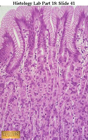 The main point of this set of slides is to look at the different specialized epithelial secretory cells of the GI tract and consider the process of breakdown and absorption of foods. This means looking primarily at the details of the GI mucosa. The first slide shows the long, straight mucosal glands of the main body of the stomach: the so-called gastric or fundic glands. The simple columnar epithelium lining the main lumen of the stomach secretes a mucoid substance (often called mucigen). This same epithelium continues on down into short "pits", which might be considered to be the ducts of these glands. Notice that there are no goblet cells in this lining; this is crucial to deciding whether you have stomach or intestines. The narrowest part of the pit, just before the main secretory cells begin, is called the "neck" and is lined with rather small, ill-defined "mucous neck cells", which secrete mucus. This is also the place where mitotic activity for replenishment of gland cells and surface epithelium takes place. Two or three secretory tubules may branch off of a single pit. In the long glands you can see granular chief cells and pink parietal cells.
 Detail of gastric (fundic) glands. The chief cells are paler, more granular looking, and tend to be basophilic at their base because of RER. Their zymogen granules are precursors of pepsin, the primary enzyme produced in the stomach. Seeming to hang off the sides of the glands are the rounded, pink parietal which have quite a clear acidophilic cytoplasm. Their secretions pass to the tubule lumen via channels between the chief cells; the lumens of the glands are too narrow to see here except perhaps in the gland at lower right center. Parietal cells secrete the precursors of HCI and the anti-pernicious anemia factor (also known as intrinsic factor). While viewing this slide, notice the loose areolar connective tissue of the lamina propria which packs around the individual glands.
 EM of chief cell, which has the features of so many other serous, protein- secreting cells of the body. The nucleus is round and lies near the base. There is a lot of RER at the base. The secretory zymogen granules lie apically, are quite large, and will be expelled into the lumen of the gland by merocrine secretion.
EM of an endocrine (or argentaffin) cell in the wall of a gastric gland. Secretory peptide granules are small and dark and leave the cell at the base, going directly to capillaries in the lamina propria. The RER of a chief cell is visible just to the left of the endocrine cell; at the upper left-hand edge of the picture there is a large, round, pale gray zymogen granule half showing in the chief cell cytoplasm. Note its size in comparison with the small granules of the endocrine cell.
 This jejunum will serve as an example of the mucosa of the small intestine. Fairly broad villi at the top are much wider than the spaces between the intestinal crypts of Lieberkuhn below. A cellular lamina propria constitutes the core of villi and the packing around crypts. Two (or perhaps three) narrow layers of muscularis mucosae lie below the glands; then the fibrous submucosa begins. Goblet cells are evident in the epithelial lining of both the villi and the crypts. Paneth cells, if present, would lie in the bases of the crypts.
Detail of base of a crypt, showing large zymogen granules in the apical region of several Paneth cells. Notice goblet cells among the simple columnar lining cells higher up in the tubule. Notice also that there is a capillary in the lamina propria below the gland.
Two more bases of crypts of Lieberkuhn. The one on the right has a couple of Paneth cells with large, acidophilic granules. The one on the left has an endocrine cell with bright orange-pink granules toward the base of the cell. Wandering eosinophils and lymphocytes are prominent among the connective tissue cells of the lamina propria.
Detail of Paneth cells with bright red secretory granules at their apices. These are the precursors of peptidase and various lysozymes. (NOTE: Paneth cells look very much like gastric chief cells in EM, with the large apical zymogen granules and a lot of basal RER.
Detail of another cross cut intestinal gland showing both: (1) Paneth cells with large, red, apical granules and (2) argentaffin (enterochromaffin) cells with small, orange-red, basal granules. The former are exocrine secreters; the latter, endocrine.
EM of enterochromaffin (endocrine) cells from a crypt. Granules lie basal to the nucleus in each case, ready to be released to the capillaries outside in the lamina propria. Notice the capillary in the lower right corner.
Villi of small intestine. Lamina propria forms their core.
The empty space in the villus is a lacteal (lymphatic capillary). Note the simple columnar epithelium covering the surface of the villus, and the endothelium lining the lacteal.
Villus with distended capillary inside (filled with pale r.b.c.'s). Bright red cells in lamina propria connective tissue are eosinophils. A goblet cell is at lower right.
Detail of lamina propria of villus, with a capillary and some longitudinal strands of smooth muscle cells to the left of the capillary. Many cells in the lamina propria are lymphocytes.
Detail of the surface epithelium of a villus, with its striated border for absorption.
Scanning EM of the surface of a villus, showing cell outlines of the columnar epithelial cells. Their fuzzy looking surface is covered with microvilli. This, of course, increases greatly the surface area available for absorption.
Detail of lining epithelium of the intestines, especially as seen covering a villus. The goblet cell is pouring out mucus. The columnar cells have a clear striated (or brush) border of microvilli. If you'll recall, these are the cells we studied much earlier for the terminal web lying just below the microvilli and for the terminal bars (dark points between cells at their top borders). The bars represent junctional complexes.
Detail of a villus stained with Mallory. The central space is a lacteal lined with endothelium. Around it is the cellular lamina propria c.t.. In the columnar epithelium of the surface of the villus several features are particularly clear: (1) The blue goblet cells discharging mucus; (2) The "terminal web" forming a thin red line at the base of the microvilli (striated border); and (3) The bright blue basement membrane separating epithelium from lamina propria. Notice several lymphocytes (round nuclei) passing through the epithelium.
EM of striated border (microvilli). It is within the mucopolysaccharide surface coat (the glycocalyx) that lactase and some other digestive enzymes become activated. The cell membrane of the microvilli extends carbohydrate "arms" which interlace to form this tangled-looking glycocalyx. |
| | | | | | صور طبية هيستولوجى - Histology | |
|
مواضيع مماثلة | |
|
| | صلاحيات هذا المنتدى: | لاتستطيع الرد على المواضيع في هذا المنتدى
| |
| |
| |